Radiological Case: Mega appendix


CASE SUMMARY
A 51-year-old man with no significant medical history presented to the emergency department with moderate diffuse and constant abdominal pain for the past 4 days. He was afebrile and his vital signs were normal. His abdomen was mildly tender diffusely with more focal pain in the right lower and upper quadrants without peritoneal signs. While in the ED, the patient was given intravenous fluids and IV toradol for pain management. Complete blood count, comprehensive metabolic panel, and urinalysis were obtained and were noncontributory.
IMAGING FINDINGS
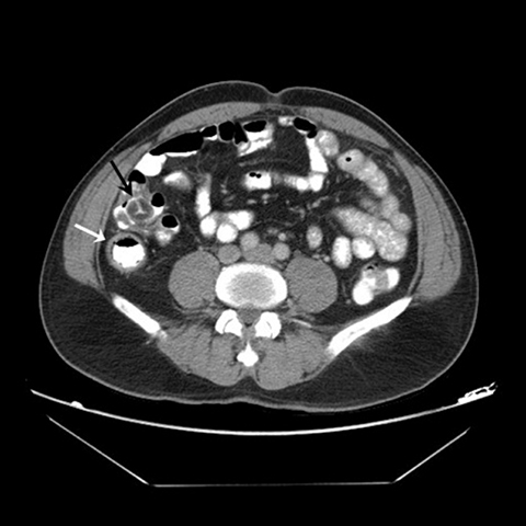
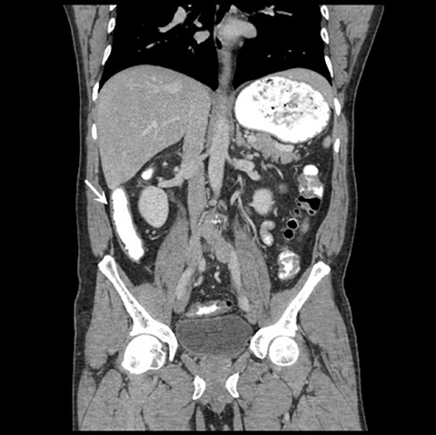
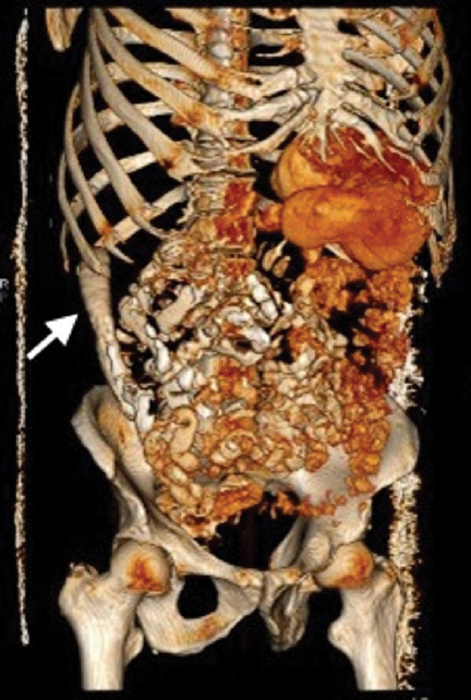
A 64-slice CT of the abdomen and pelvis with IV and PO contrast was performed with multi-planar reconstructions (Figures 1-3). The exam revealed a markedly enlarged, blind-ending, retrocecal tubular loop of bowel that entirely filled with oral contrast, extending to the subhepatic region, consistent with the appendix. It measured 2.5 cm wide and 13 cm in craniocaudal dimension. Although its wall was slightly prominent, there were no surrounding inflammatory changes and there was no appendicolith. The patient’s symptoms improved after IV fluids and Toradol were administered. After discussion with the on-call surgeon, the patient was referred for outpatient follow-up and discharged. The patient was contacted as an outpatient and indicated his symptoms resolved completely without recurrence.
DIAGNOSIS
Mega appendix
DISCUSSION
The vermiform appendix is a continuation of the cecum of the large intestine. The appendix ranges in length from 5-10 cm and 0.5-1 cm in width.1 The annual rate of acute appendicitis was 9.38 per 10,000 in 2008.2 CT imaging has been reported to be 91-98% sensitive and 75-93% specific for the diagnosis of appendicitis.3 CT is commonly the first line approach to abdominal pain, with reported accuracy rates up to 95-100% for the diagnosis of appendicitis.4 Prior to appendectomy, preoperative CT use was 94.7% in 2007, an increase from 18.5% in 1998.5 As a result, it is imperative for the radiologist to understand atypical anatomical variations of the appendix.
The appendix position can vary in relation to the other abdominal organs.6 The base is usually located about 2 cm below the ileocecal valve (Figure 1).1 However, the free end of the appendix can occupy a variety of positions in relation to the small and large bowel: anterior, medial, lateral, inferior, superior, superiorlateral and retrocolic.6 The average length of an appendix in an adult is 9.5 cm.7 Tamburrini et al evaluated the CT images of 372 patients. The appendix was visualized in 305/372 of the abdominopelvic scans. The average appendix diameter range was between 3-10 mm and wall thickness was 1.5 mm.6 Our patient had an appendix that was much wider and longer than the average appendix.
Large appendixes have been reported in the medical literature, mostly outside of radiology. One of the first was reported in 1890 by F. Grauer. He discovered a 33-cm-long appendix in a cadaver.8 The next largest appendixes were discovered incidentally from autopsy specimens, 21.5, 22, 23 and 24 cm in length.8,9 In 1920, Lake reported the case of a 22-year-old male patient with chronic abdominal pain who presented with acute appendicitis with a perforated tip. He underwent an appendectomy, which revealed a 29.4 cm appendix.10 In 1932, Collins performed a study evaluating 4,680 appendix specimens and discovered the longest appendixes found on autopsies of men who died from conditions unrelated to the appendix; one was a 28-cm appendix found in a 40-year-old man and the other was a 24.5 cm appendix found in a 76-year-old male.9 From 1923 to 1963, Collins performed a large study of 71,000 appendix specimens, 91% of which were from appendectomy and 9% from post-mortem evaluation. The Collins study was one of the largest studies to evaluate appendixes but in the final report there was no assessment of the variability in size of the appendix.11
In 2005, a case report of a 36-year- old man with a 28 cm, torsed and necrotic appendix was reported.12 A case report from the United Kingdom in 2009 described a 10-year-old patient with a 17-cm-long inflamed appendix found during appendectomy. The tip of the appendix had reached the subhepatic area.7 In 2011, a 25-year-old man was found to have a 20-cm, noninflamed appendix adhering to his inguinal sac. He underwent a mesh plug hernia repair with an appendectomy.13 The most recent reported case was in 2013 in India, where a 28 cm appendix was found during routine dissection in a medical school cadaver laboratory.14
The previous case reports focus on cases of large appendixes found on surgery or at autopsy. The vast majority lack diagnostic imaging results. The classic CT findings of nonperforated acute appendicitis include appendiceal wall thickening and dilatation, periappendiceal inflammatory changes, wall hyperemia, and nonfilling with enteric contrast.15 The appendix presented in our case report filled entirely with oral contrast and lacked surrounding inflammatory changes, thereby excluding the diagnosis of acute appendicitis despite its unusually large dimension. That the patient’s pain resolved after conservative management is also in keeping with the lack of acute appendicitis. Furthermore, previous radiological literature has described the importance of recognizing atypical anatomical variants on CT scans of the appendix in order to appropriately diagnose appendicitis. Various pitfalls have been discussed, including variable appendiceal location, congenital abnormalities (such as malrotation), and the interference of coexisting pathologies.16 However, variability in size of the appendix and its role in accurate diagnosis of appendicitis has not been explored to the same degree. Therefore, our case depicts the importance of recognizing that a markedly enlarged appendix may be a normal anatomic variant.
CONCLUSION
The myriad appearances of the appendix can make diagnosis of acute appendicitis challenging. Our case is unique compared to other prior case reports as our patient is a living, healthy, middle-age patient with an extraordinarily large, non-inflamed appendix. Therefore, this unusually wide mega appendix is felt to be a normal anatomic variant. Familiarity with such an atypical appearance can allow for a more accurate diagnostic approach, help prevent the erroneous diagnosis of acute appendicitis, and help avoid unnecessary procedures.
REFERENCES
- Schwartz SI, Shires GT, Spencer FC. Principles of surgery. 5th ed. New York: McGraw-Hill; 1969: 1315.
- Buckius MT, McGrath B, Monk J, et al. Changing epidemiology of acute appendicitis in the United States: Study period 1993–2008. J Surg Res. 2012; 175:2: 185-190.
- Levine CD, Aizenstein O, Wachsberg RH. Pitfalls in the CT diagnosis of appendicitis. Brit J Radiol. 2004; 77:921:792-799.
- Rao PM, Rhea JT, Novelline RA, et al. Helical CT technique for the diagnosis of appendicitis: Prospective evaluation of a focused appendix CT examination. Radiology. 1997; 202:1:139-144.
- Coursey CA, Nelson RC, Patel MB, et al. Making the diagnosis of acute appendicitis: Do more preoperative CT scans mean fewer negative appendectomies? A 10-year study. Radiology. 2010; 254:2:460-468.
- Tamburrini S, Brunetti A, Brown M, et al. CT appearance of the normal appendix in adults. Eur Radiol. 2005;15:10: 2096-2103.
- Alzaraa A, Sunil Chaudhry. An unusually long appendix in a child: A case report. Cases Journal. 2009;2:1: 7398.
- Kelly HA, Hurdon E. The vermiform appendix and its diseases. Philadelphia, PA: WB Saunders; 1905:136.
- Collins DC. The length and position of the vermiform appendix: A study of 4,680 specimens. Ann Surg. 1932; 96:6:1044.
- Lake GB. Report of an extremely long vermiform appendix. JAMA. 1920; 75:19: 1269-1269.
- Collins DC. 71000 human appendix specimens. A final report, summarizing forty years’ study. Am J Proctol. 1963;14: 365-381.
- Takuya Y, Yoshikazu M, Yojiro K, et al. A case report of torsion of an unusually large vermiform appendix. J Japan Surg Assoc.2005;6;1376-1378.
- Psarras K, Lalountas M, Baltatzis M, et al. Amyand’s hernia-a vermiform appendix presenting in an inguinal hernia: A case series. J Med Case Rep. 2011; 5:1: 463.
- Boddeti RK, Kulkarni R, Murudkar PK. Unique 28cm long vermiform appendix. Int J Anat Res. 2013; 1:2: 111-14.
- Leite, NP, Pereira JM, Cunha R, et al. CT evaluation of appendicitis and its complications: Imaging techniques and key diagnostic findings. Am J Roentgenol. 2005;185:2:406-417.
- Shademan A, Tappouni RF. Pitfalls in CT diagnosis of appendicitis: Pictorial essay. J Med Imag Rad Onc. 2013; 57:3: 329-336.
