Chronic recurrent multifocal osteomyelitis
Chronic recurrent multifocal osteomyelitis
Findings
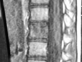
The sagittal T1 series of the thoracolumbar spine showed mild superior end-plate compression of the T12 vertebral body with abnormal marrow signal, initially presumed to be post-traumatic (Figure 1).
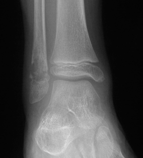
Plain film and coronal T1 (450/14) images of the right ankle (Figure 2) demonstrated an osteolytic lesion in the right distal fibular metaphysis. Coronal FSE IR (5400/63) showed a right proximal tibial lesion (Figure 2). Given the concern for multifocal bacterial osteomyelitis and malignancy, biopsy of the distal fibular metaphyseal lesion was performed, revealing both acute and chronic osteomyelitis. All bacterial and fungal cultures were negative. A bone marrow biopsy concurrently performed at this time showed no findings of hematopoetic malignancy. Conservative treatment was given, with the presumptive diagnosis of chronic recurrent multifocal osteomyelitis.
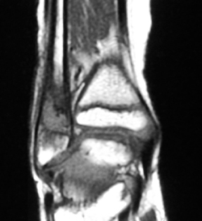
Coronal FSE IR (5400/63) of the lower extremity, taken 15 months later, showed new symmetric lesions in the distal tibial metaphyses, as well as lesions in the left proximal tibial metaphysis (Figure 3). The previously noted right proximal tibial metaphyseal lesion had resolved. The patient was again treated conservatively with resolution of symptoms.
Discussion
Chronic recurrent multifocal osteomyelitis (CRMO) is an inflammatory disease of bone that occurs primarily in children and adolescents.1 Though it generally has a benign, self-limited clinical course, its variable clinical findings can often mimic more serious diseases such as bacterial osteomyelitis and malignancy.2 Recognition of the imaging findings of this protean condition plays a crucial role in its diagnosis.
Symptoms may include low-grade fever and malaise, with local pain and swelling of the affected bones, although this constellation of findings is not consistently present.3,4 Laboratory findings are relatively few and point to nonspecific inflammation, with elevation of the erythrocyte sedimentation rate (ESR). Microbiologic cultures of the blood are negative.5
The most characteristic features are recurrent episodes of active disease followed by remission. Reactivation occurs either in previously involved osseous locations or in different sites.1 The most common locations are the metaphyses of long bones and clavicles. Less frequent involvement is seen in the vertebrae and pelvis, while other locations are uncommon.6 Symmetric lesions have been classically described, though this is an inconsistent finding.7
The radiographic findings appear similar to acute bacterial osteomyelitis. Metaphyseal lesions are osteolytic, with a thin sclerotic rim often seen in healing lesions.8 The degree of periosteal reaction can vary, as can the presence of soft tissue swelling.9 Bone scintigraphy is useful in identifying the presence of multifocal disease, which helps in making the correct diagnosis.
MR imaging is especially advantageous in detecting early disease before radiographic findings are present. In addition to demonstrating the characteristic findings of marrow edema, it is useful in excluding stigmata associated with bacterial osteomyelitis such as soft tissue abscesses, sinus tracts and sequestra.10
Conclusion
Though chronic recurrent multifocal osteomyelitis is a diagnosis of exclusion, the clinical course, presence of multiple reactivating and remitting lesions over time, and absence of positive microbiologic results should suggest the diagnosis. As the disease is self-limiting, knowledge of its characteristic appearance can prevent overly aggressive medical and surgical evaluation and treatment.
- Jurriaans E, Singh NP, Finlay K, Friedman L. Imaging of chronic recurrent multifocal osteomyelitis. Radiol Clin North Am. 2001;39:305–327.
- Chow LTC, Griffith Jf, Kumta SM, Leung PC. Chronic recurrent multifocal osteomyelitis: A great clinical and radiologic mimic in need of recognition by the pathologist. APMIS. 1999;107:369–379.
- Demharter J, Bohndorf K, Michl W, Vogt H. Chronic recurrent multifocal osteomyelitis: A radiological and clinical investigation of five cases. Skeletal Radiol. 1997;26:579–588.
- Gamble JG, Rinsky LA. Chronic recurrent multifocal osteomyelitis: A distinct clinical entity. J Pediatr Orthop. 1986;6:579–584, .
- Handrick W, Hörman D, Voppmann A, et al. Chronic recurrent multifocal osteomyelitis: Report of eight patients. Pediatr Surg Int. 1988;14:195–198, .
- Jurik AG. Chronic recurrent multifocal osteomyelitis. Semin Musculoskelet Radiol. 2004;8:243–253.
- Wiener MD, Newbold RG, Merten DF. Chronic recurrent multifocal osteomyelitis (case report). AJR Am J Roentgenol. 1986;147:87–88.
- Rosenberg ZS, Shankman S, Klein M,Lehman W. Chronic recurrent multifocal osteomyelitis. AJR Am J Roentgenol. 1988;151:142–44.
- Carr AJ, Cole WG, Roberton DM, Chow CW. Chronic multifocal osteomyelitis. J Bone Joint Surg Br. 1993;75:582–591.
- Jurik AG, Egund N. MRI in chronic recurrent multifocal osteomyelitis. Skeletal Radiol. 1997;26:230–238.