Radiological Case: Perched facet
SUMMARY
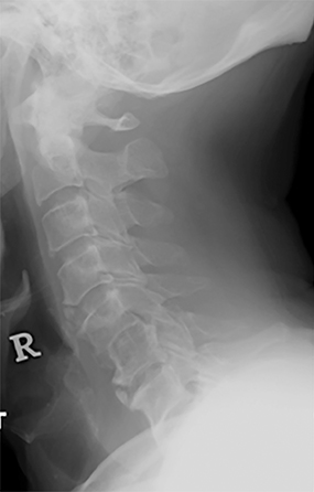
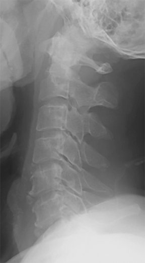
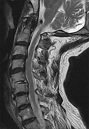
A 71-year-old male presented after a vehicular collision complaining of severe neck pain without focal weakness or numbness. The patient was wearing a seatbelt during the impact and reported hyperflexion of his neck upon impact. No neurological deficits were elicited on clinical examination. Initial cervical spine radiographs were performed at the emergency department (Figure 1) and compared with his previous study done a year ago (Figure 1). Noting the radiographic findings, a magnetic resonance (MR) image was promptly performed (Figure 2).
IMAGING FINDINGS
Figure 1A shows a lateral cervical radiograph done at first presentation, which showed interval development of mild anterior subluxation of C5 over C6 (involving approximately 25% of the vertebral width), reduced C5-6 intervertebral disc space, disruption, and distraction of the normal facet joint alignment and widening of the interspinous distance; these were new findings in relation to a prior study performed for cervical spondylosis one year ago (Figure 1B).
Figure 2 is a sagittal T2WI MR showing focal kyphosis and anterior subluxation at C5-6 consistent with hyperflexion injury. There is high T2W fluid-signal in the disc consistent with acute disc injury. No significant oedema in the posterior longitudinal ligament at this level is detected. There was no evidence of cord oedema or injury to the interspinous ligaments. Background cervical spondylosis and multi-level osteochondral bars are observed.
DIAGNOSIS
Left-sided (unilateral) perched facet
DISCUSSION
Unilateral facet joint injuries are uncommon and represent approximately 6% of all cervical spine injuries. The spectrum of injuries to the facet joint encompasses fracture, subluxation, and dislocation with descriptors of locked, jumped or perched facet, with associated disco-ligamentous injury.1,2
Unilateral injury to the facet joint is generally thought to cause a certain degree of rotational instability as the injured facet and lateral mass complex rotates around the intact contralateral facet. There is also an association with concurrent disco-ligamentous soft tissue injuries secondary to distraction, especially involving the posterior annulus of the disc, posterior longitudinal ligament and the interspinous ligaments, reflecting its hyperflexion injury mechanism.3,4
Radiographic signs on lateral projection include translation of the vertebral body up to 25% of the anteroposterior diameter of the adjacent vertebral body, focal kyphosis, reduced overlap of the articular processes and fanning or widening of the interspinous distance. Displacement of the interfacetal joint with rotational deformity may also produce a “double-appearance” of articular facets above the level of injury reminiscent of a “bow tie”. This appearance results from the displacement of the articular mass anteriorly at the level of the injury with respect to the contralateral side. The “bow tie” sign was however, only noted in about one-third of cases of unilateral facet joint dislocation.2
MRI is important for the evaluation of concurrent disco-ligamentous and cord injury and findings on fluid-sensitive sequences include the presence of prevertebral and ligamentous edema, marrow edema at the site of fractures and increased cord signal indicating contusion. Cord hemorrhage may be evaluated with T2* GRE sequence.
Treatment of unilateral facet joint injuries depends on the degree of bony, ligamentous and cord involvement as well as spinal stability and may be non-operative for simple stable unilateral facet dislocation, as in our patient, (immobilization or closed reduction) or surgical (failed conservative measures, persistent instability, signs of middle column disruption at the outset, cord injury or epidural haematoma requiring surgical decompression).
CONCLUSION
Unilateral cervical facet-joint injury, though uncommon by itself, should be suspected in patients involved in road traffic accidents and sports injuries with a hyperflexion/rotation mechanism. It can be diagnosed on plain radiographs and is important to recognize and further evaluate for associated soft tissue injuries to avoid further instability or delayed neurological deficits.
REFERENCES
- Marcel F et al. Clinical outcomes of 90 isolated unilateral facet fractures, subluxations, and dislocations treated surgically and nonoperatively. Spine. 2007;32(26):3007-3013.
- Young JW, Resnik CS, DeCandido P, Mirvis SE. The laminar space in the diagnosis of rotational flexion injuries of the cervical spine. AJR Am J Roentgenol. 1989;152(1):103-107.
- Laporte C et al: Severe hyperflexion sprains of the lower cervical spine in adults. Clin Orthop. 1999;(363):126-134.
- Fazl M et al: Posttraumatic ligamentous disruption of the cervical spine, an easily overlooked diagnosis: Presentation of three cases. Neurosurgery. 1990;26(4):674-678.