Too Much To Swallow: Imaging to Detect Contraband in the Gastrointestinal Tract


CASE SUMMARY
A 21-year-old was brought to the emergency department in police custody with stable vital signs and no complaints. They demonstrated guaiac-positive stool and a palpable object on rectal examination. Two days prior, they arrived in the United States from another country, where they report ingesting 100 bags of cocaine. From time of custody to hospital visit, the patient passed 87 bags. Poison control recommended performing CT of the abdomen/pelvis. An additional 3 bags passed in the hospital prior to imaging. Subsequent to imaging, enemas were administered, facilitating uncomplicated passage of the remaining 10 bags.
IMAGING FINDINGS
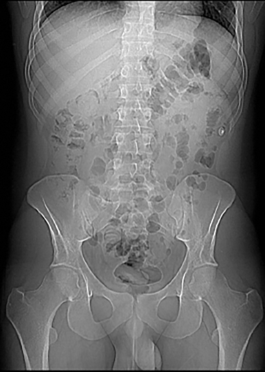
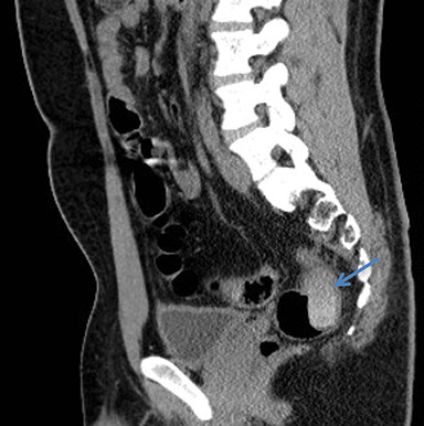
Abdominal radiography failed to reveal foreign bodies (Figure 1). Noncontrast CT of the abdomen and pelvis revealed approximately 10 radiodense cylindrical opacities scattered throughout the gastrointestinal tract, including the stomach, and was without evidence of intestinal perforation (Figure 2).
DIAGNOSIS
Foreign-body contraband in the GI tract
DISCUSSION
Foreign body ingestion may be accidental or intentional. Accidental foreign body ingestion is most commonly seen in children, whereas adults are more likely to perpetrate intentional ingestion and rarely as an isolated incident.1 A specific type of foreign body ingestion, termed “body packing,” describes the illegal intra-corporeal concealment of containers or packets containing any number of illicit substances (most commonly cocaine, heroin, or cannabis) which may already be diluted or adulterated with other pharmacologically active or inactive substances for enhanced effect and/or increased profit on resale.2-5
“Body packing” was first reported in 1973, when a 21-year-old patient who swallowed a condom of hashish presented with small-bowel obstruction.2 Since then, the practice has become more widely recognized: patients ingest containers, or insert them rectally, vaginally, or even in the external auditory canal.2,6
Although 80-90% of ingested foreign bodies pass spontaneously, patients are at risk of serious medical complications, including bowel obstruction, intestinal perforation and peritonitis, respiratory distress due to packet aspiration, or packet rupture leading to acute drug toxicity, overdose, and death.2, 7 In asymptomatic patients, imaging is essential for identifying drug-containing foreign bodies, their number, and location; in symptomatic patients, it becomes crucial for detecting medical complications.2
With a sensitivity of 60-90%, abdominal radiography is commonly used for foreign body detection as it is widely available and can be performed relatively quickly and easily.4 A number of radiographic signs are suggestive of body packing. A drug package surrounded by a radio-transparent halo (due to air-trapping between layers of wrapping) is termed “double condom sign.” The “parallelism sign” occurs when the packages align in parallel along the course of the gastrointestinal lumen.4 The “Tic Tac sign” and “bag-of-eggs sign,” both highly specific for solid forms of drugs, look as they sound: drug packets appear as smooth spherical to cylindrical radiopaque structures within the intestinal lumen.4, 8 The “multiplicity sign” describes numerous, well-defined opacities within the gastrointestinal tract inconsistent with normal gut contents.7 “Rosette sign” describes radiolucent areas of air-trapping in the knots of tied-off balloons or condoms. This is less frequently seen currently due to the general transition from manual wrapping to the current trend of wrapping via more industrial methods).4 Physicians should also be aware of strategies employed to reduce the ability of radiographic imaging to detect contraband (eg, packaging with carbon paper or aluminum foil, which decrease radiodensity);4 as such, high clinical suspicion should not necessarily be disregarded in the presence of an apparently negative abdominal X-ray.
The sensitivity of ultrasonography is lower than that of radiography; in detecting ingested packets, ultrasound demonstrates a higher rate of both false-positives and false-negatives owing to its inability to penetrate bowel gas,4, 9 and cannot identify the number of packets or allow for any determination of their contents.10 If encountered, however, an ingested packet typically appears as a linear or ovoid hyperechoic structure with posterior acoustic shadowing.4, 9 Despite being noninvasive, inexpensive, and free from ionizing radiation, ultrasound is highly user-dependent and, therefore, unlikely to be widely adopted for screening.4, 9 In the case that radiographic or sonographic studies are negative or unequivocal, CT scanning should follow.4
Abdominal CT is becoming the diagnostic imaging modality of choice for detecting drug-containing foreign bodies, owing to its high contrast resolution and superior sensitivity and specificity to X-ray for detecting both liquid and solid drug-filled packets.4, 9, 11 The primary deterrent to using CT for initial screening, however, is the radiation exposure associated with CT.10 CT is also instrumental in identifying complications of body packing, including packet rupture, intestinal obstruction, and intestinal perforation.4 Recent literature discourages administration of oral or rectal contrast due the potential to obscure the packages due to similarities in density.4 Using a lung window setting to view the abdominal CT scan can augment packet detection, and classic radiographic signs of body packing, such as the double condom sign, parallelism, and Tic Tac sign can also be identified with CT.8 The measurement of Hounsfield units (HU) may assist in identification of the smuggled substance; experiments have shown that cannabis has a density similar to bone (700 HU), cocaine is less dense than fat (-219 HU), and heroin even more hypodense (-520 HU). The presence of cutting agents, however, can derange these densities significantly.8
Magnetic resonance imaging (MRI) remains underexplored for identifying body-packers; regardless, MRI is rarely utilized because of its costliness, limited availability, and requirements of high-volume oral contrast, along with spasmolytic agents, to reduce artifacts caused by peristalsis.10, 12
CONCLUSION
The radiologist’s ability to identify foreign bodies in asymptomatic patients is likely to become increasingly useful to law enforcement, especially in an age when body-packing is becoming more common and drug traffickers are becoming more creative. Imaging will also continue to be vital for detecting medical complications of the practice.
REFERENCES
- Palese C, Al-Kawas FH. Repeat Intentional Foreign Body Ingestion: The Importance of a Multidisciplinary Approach. Gastroenterology & Hepatology. 2012;8:485-486.
- Pinto A, Reginelli A, Pinto F, et al. Radiological and practical aspects of body packing. Br J Radiol. 2014;87:20130500.
- Cole C, Jones L, McVeigh J, et al., CUT: A guide to adulterants, bulking agents and other contaminants found in illicit drugs. Liverpool: Liverpool John Moores University, 2010.
- Cappelletti S, Piacentino D, Sani G, et al. Systematic review of the toxicological and radiological features of body packing. Int J Legal Med. 2016;130:693-709.
- Poletti PA, Canel L, Becker CD, et al. Screening of illegal intracorporeal containers (“body packing”): is abdominal radiography sufficiently accurate? A comparative study with low-dose CT. Radiology. 2012;265:772-779.
- Esterson YB, Patel V, Nicastro J, Friedman B. Plain radiography may underestimate the burden of body packer ingestion: A case report. Clin Imaging. 2017;44:57-60.
- Ray A, Nayan A, Katariya K, Sharma SK. Body Packer Syndrome: A Radiological Denouement! The Journal of Emergency Medicine. 2018.
- Kai Patrick Tsang H, Kei Kathy Wong C, Fung Wong O, et al. Radiological features of body packers: An experience from a regional accident and emergency department in close proximity to the Hong Kong International Airport. Hong Kong J Emerg Med. 2018:102490791877008.
- Berger FH, Nieboer KH, Goh GS, Pinto A, Scaglione M. Body packing: a review of general background, clinical and imaging aspects. Radiol Med. 2015;120:118-132.
- Sica G, Guida F, Bocchini G, et al. Imaging of drug smuggling by body packing. Semin Ultrasound CT MR. 2015;36:39-47.
- Bulakci M, Kalelioglu T, Bulakci BB, Kiris A. Comparison of diagnostic value of multidetector computed tomography and X-ray in the detection of body packing. Eur J Radiol. 2013;82:1248-1254.
- Reginelli A, Russo A, Urraro F, et al. Imaging of body packing: errors and medico-legal issues. Abdom Imaging. 2015;40:2127-2142.
Affiliations: St. George’s University School of Medicine, True Blue, Grenada (Ms. Solomon) is with St. George’s University School of Medicine, True Blue, Grenada; Department of Radiology, Queens Hospital Center, Icahn School of Medicine, Mount Sinai, Jamaica, NY (Dr Singh Tuli).