UC San Diego Gets NIH Grant to Develop Whole-Body Inflammation Imaging
Researchers at University of California San Diego School of Medicine have been awarded two new grants by the National Institutes of Health (NIH), totaling $6.7 million, to develop and clinically test technologies that can noninvasively examine and quantify immune cells found in tumors. These immune cells, called macrophages, are involved in the body’s normal inflammatory responses, but they also make up a significant portion of solid tumors. The density of macrophages in a tumor can affect how it responds to treatment, so the ability to count them noninvasively could help doctors decide which therapies will be most effective.
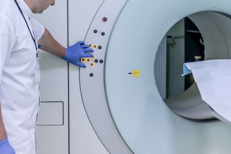
The technology works by feeding macrophages tiny drops of a dye that can be detected by magnetic resonance imaging (MRI). The researchers will test the technology, called TAM-Sense, in patients with recurrent head and neck tumors. The new phase I clinical study will be the first time TAM-Sense is being tested in patients. The team will also adapt the technology to be used with positron emission tomography (PET) to enable whole-body imaging.
“Visualizing a patient’s inflammatory sites throughout the body will be invaluable for accurate clinical diagnosis and for planning precise therapeutic interventions,” said Eric Ahrens, PhD, a professor in the Department of Radiology at UC San Diego School of Medicine. “Current approaches using biopsies are invasive, and some tumors are inaccessible to biopsy. There is an urgent need for new, whole-body imaging technologies.”
The TAM-Sense agent is a fluid containing extremely small drops of a biologically inert fluorocarbon dye, which is dispersed into the bloodstream when the agent is injected. Macrophages detect and engulf these droplets, then accumulate at sites of inflammation. To detect the dye inside the macrophages, the method uses a specially modified MRI scanner. For the patient, the experience is similar to a standard MRI scan.
Beyond its use for cancer, TAM-Sense could also have broad clinical applications for other diseases that have substantial inflammatory components, such as autoimmune disorders, cardiovascular diseases and infectious diseases. Macrophages are often found at sites of pain in the body, and imaging tools that can pinpoint the anatomical location of these sites will enable more precise pain management.
In addition to testing TAM-Sense, the researchers will also further enhance its capabilities by making it compatible with PET scans. Unlike MRI, which provides high resolution images over limited fields of view, PET excels at full-body scanning for disease detection. While the PET approach will require further preclinical testing before it is ready for use on patients, the researchers see this as the next step for the technology.
“Right now, we’re testing one iteration of the technology in patients but there’s also a bigger story at play in terms of empowering MRI and PET with unprecedented precision, which could have wide-reaching implications for diagnostics as a whole,” said Ahrens. “This is only the first step.”