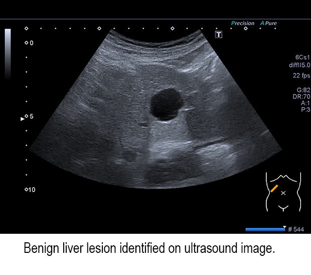
Ultrasound and benign focal liver lesions
The capabilities and performance of the latest generation of ultrasound equipment have improved the ability to discover focal liver lesions during abdominal exams, and for this reason, the number being identified has increased markedly. In addition to being able to identify malignant lesions, radiologists also need to distinguish the types of benign lesions accurately and efficiently to determine what treatment is needed, if any.
Radiologists can benefit by having a fundamental knowledge of the prevalence and image morphology of benign lesions. For this reason, a multidisciplinary team of German researchers conducted a study of over 45,000 patients at the University Hospital Ulm who had an ultrasound exam during a 10 year period for the diagnosis of benign focal liver lesions. In an article published in the January issue of Abdominal Radiology, they compared their findings with 10 similar peer-review journal published studies. For the most part, their findings were comparable. Their findings are intended to be a reference for other radiologists.
 A total of 15.1%, or 6851 patients, had at least one focal liver lesion of interest. These patients represented a mix of 55.1% inpatients and 44.9% outpatients. Focal fatty sparing was identified in 2839 cases, or 41.4% of the patients with an identified lesion of interest. The mean maximum measured size of the focal fatty was 20.6 mm. Focal fatty sparing was diagnosed most frequently in patients, predominantly men, between 51 and 60 years of age. However, if a patient had hepatic steatosis, 90% of focal fatty sparing was identified in patients under the age of 30.
Hepatic cysts were the second most commonly identified benign liver lesion, at 38.4% of patients with at least one focal liver lesion. This type of lesion is age specific, with only 21 out of 2,631 patients under the age of 30 and 1,012 in patients over the age of 70.
There were 1,640 cases of hepatic hemangiomas, or 23.9% of patients with a focal liver lesion of interest. Age-specific prevalence was relatively uniform with the largest percentage in patients between 51 and 60 years of age. The smallest percentage was 7% for the age group under 30 years. Over three quarters of the hemangiomas diagnosed were solitary, with an average size of 20.1 mm.
Focal nodular hyperplasia (FHN) and hepatic adenomas were rare, with 81 and 19 cases respectively diagnosed. Four fifths of the FHNs were found in women, with almost 65% of the total in patients under the age of 40. Prevalence fell with increasing age; only seven patients over the age of 61 were diagnosed. FHNs were the largest fatty focal lesions, with an average size of 51.6 mm.
Hepatic adenomas were solitary in 90% of the cases, and had a mean size of 39.0 mm. Sixteen of the 19 cases were identified in women, the largest group found in patients aged 41-50 years.
Lead author Tanja Eva-Maria Kaltenbach, MD, of the department of internal medicine, and colleagues, summarized their research by saying “the first possibility to be considered on the incidental discovery of space-occupying lesions of the liver – especially if hepatic steatosis is present – is focal fatty sparing. Simple hepatic cysts and hemangiomas are the most common focal liver lesions. The finding of a FNH or an adenoma is rarely a fortuitous result.”
REFERENCE
- Kaltenbach TE, Engler P, Krazer W, et al. Prevalence of benign focal liver lesions: Ultrasound investigation of 45,319 hospital patients. Abdom Radiol. 2016 41;1:25-32.
