Vereos PET/CT: Leveraging Digital Precision for Innovative Approaches
Supplement to November-December 2019 Applied Radiology. Sponsored by Philips Healthcare.
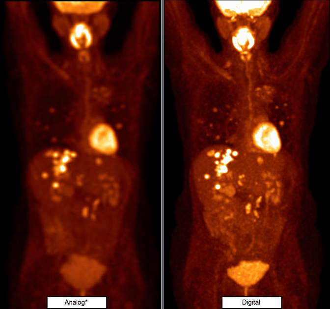
Frequently used in oncology, cardiology and other clinical areas, PET/CT has become an indispensable diagnostic and prognostic imaging tool in health care. Contributing to this growth of utilization of PET imaging are such advances as the digital detector technology in Philips’ Vereos PET/CT. Digital detectors improve sensitivity and quantification, allowing for higher-definition imaging and detection of smaller lesions.
Leveraging these advancements and commercial availability, work is now under way to further optimize image quality, low dose protocols, and image acquisition times. The Vereos digital PET/CT, for example, is being used at the Wright Center of Innovation in Biomedical Imaging at Ohio State University’s Wexner Medical Center in several initiatives aimed at achieving high-quality images at low doses and fast acquisition times.
A digital future
“There’s really no question anymore that digital PET is the future,” says Michael V. Knopp, MD, Novartis Chair of Imaging Research and Director of the Wright Center of Innovation. “What we’re doing now is applying the benefits of the different technological advancements of digital technology. One of the areas we’re focused on is improving image quality while imaging faster and at a low tracer dose, which is very relevant for whole body imaging, but also for specific disease applications such as prostate, and neuro and cardiac imaging.”
Dr. Knopp and his team at the Wright Center collaborated with Philips on the initial evaluation of the digital PET technology in 2014. This partnership led to the commercialization of this innovation by Philips as a Vereos PET/CT system. The Wright Center has been using the Vereos ever since. Dr. Knopp retains a keen interest in optimizing PET/CT capabilities to produce high-quality images at the lowest possible radiation doses and fastest possible acquisition times.
The average speed for whole-body PET imaging is about 90 seconds per bed position using traditional PET/CT, with a whole-body acquisition averaging ~20 minutes table time. In Phase II clinical trial findings released by Dr. Knopp at the 2018 Annual Meeting of the Radiological Society of North America (RSNA),1 the investigators concluded that, using the Vereos system, ultra-fast whole-body PET/CT scans are achievable at one-tenth of the time — in just two minutes — and that consistent acceptable image quality was achievable with Vereos’ proprietary digital photon counting detector technology. The ultra-fast, two-minute whole-body PET/CT scans acquired with digital detector technology also presented with substantially fewer motion artifacts than traditional PET/CT images, the researchers found.
In addition to improving visual and quantitative quality, Dr. Knopp indicates that digital PET/CT enables the use of significantly lower tracer dose in clinical imaging.
Improving speeds and effecting change
Dr. Knopp’s work supports the possibility of PET/CT imaging at a temporal resolution improvement factor of ten and more. With this increased acquisition speed, Dr. Knopp is able to implement new approaches for dynamic imaging.
“We have taken all three of these components — image quality, low dose, and acquisition speed — and continued to evolve into more dynamic PET imaging, where we are reconstructing the imaging at a very high temporal resolution to look at the accumulation kinetics of PET tracers on target tissue,” he explains.
The ability to visualize the kinetic process has always been a hallmark of PET imaging, but according to Dr. Knopp, clinicians did not have these capabilities available anymore when whole-body static imaging with PET/CT was initially introduced.”
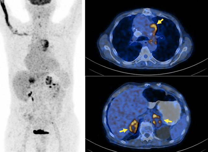
“PET can provide a lot more insight into the pathophysiological process and is so important in our molecular understanding of disease and in the molecular targets we are pursuing,” he says. “Because of the high quality and the precision of the counts we’re getting with the digital photon counting PET/CT technology, we can get a very precise determination of the PET activity within the patient.”
- Knopp MV, Knopp MI, Porter T, Binzel K, Zhang J, Giesel F, Wright C. Ultra-Fast Whole-body PET/CT Enabled by Digital Photon Counting PET Detector Technology: Findings from a Phase II Clinical Trial. Radiological Society of North America 2018 Scientific Assembly and Annual Meeting, November 25 - November 30, 2018, Chicago IL. archive.rsna.org/2018/18020222.html. Accessed September 30, 2019.