CT may Help Identify Candidates for Sublobar Resection in Early-Stage Non-Small Cell Lung Cancer
Features found on CT images may help identify which patients with stage IA non-small cell lung cancer are optimal candidates for sublobar resection, rather than more extensive surgery, according to an article in the American Journal of Roentgenology (AJR).
This retrospective study included 904 patients (453 men, 451 women; mean age, 62 years) who underwent lobectomy (n=574) or sublobar resection (n=330) for stage IA non-small cell lung cancer. Two thoracic radiologists independently evaluated findings on preoperative chest CT, later resolving any discrepancies. Recurrences were identified via medical record review.
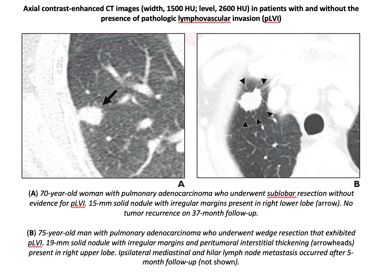
“In patients with stage IA non-small cell lung cancer, pathologic lymphovascular invasion was observed only in solid-dominant part solid nodules and solid nodules with solid portion diameter over 10 mm,” concluded corresponding author Mi Young Kim from the department of radiology at the University of Ulsan College of Medicine, Asan Medical Center.
“Among such nodules,” the authors of this AJR article continued, “peritumoral interstitial thickening (odds ratio=13.22) and pleural contact (odds ratio=2.45) were independently associated with pathologic lymphovascular invasion.” Moreover, models incorporating these features independently predicted recurrence-free survival after sublobar resection (hazard ratio=5.37–6.05).
Related Articles
Citation
CT may Help Identify Candidates for Sublobar Resection in Early-Stage Non-Small Cell Lung Cancer. Appl Radiol.
May 14, 2021