Deep Learning Reconstruction for Ultra Low-dose Lung Screening CT Finds More Nodules
 A prospective study comparing a widely used adaptive statistical iterative reconstruction-V (ASIR-V) to a deep-learning image reconstruction (DLIR) in ultra-low-dose CT lung screening reports that DLIR increased nodule detection, reduced image noise and improved measurement accuracy.
The study published in Radiology from Shanghai General Hospital found that DLIR enabled the use of ultra-low-dose chest CT, between 0.07-0.14 mSv, which is comparable to a single chest radiograph. Currently in lung cancer screening, typical low dose CT ranges from 1 mSv to 2 mSv. While iterative reconstruction improves image quality and reduces image noise, using it at doses similar to chest CT impacts the visualization of subtle imaging features.
From April to June 2020, 100 participants were scanned at 0.07 mSv and 103 were scanned at 0.14 mSv. All participants also underwent a contrast-enhanced CT. Obese patients with BMI higher than 30 kg/m2 were excluded.
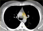
All images were processed with filtered back projection (FBP), ASIR-V and DLIR. Nodule detection rates were 75.8% (808 of 1066) with DLIR and 73.3% for ASIR-V. DLIR also reduced background image noise by 21% compared to ASIR-V. DLIR also had the most abundant subtle features, an overall malignant feature detection rate of 81.5%, improved nodule measurement accuracy and displayed more detailed malignancy-related imaging features of nodules.
The authors conclude that while it is feasible to use ultra-low-dose CT lung cancer screening of 0.14 mSv using the DLIR algorithm in underweight and normal weight participants, more studies are needed to examine clinical application and image optimization of DLIR.
Related Articles
Citation
Deep Learning Reconstruction for Ultra Low-dose Lung Screening CT Finds More Nodules. Appl Radiol.
January 20, 2022