Invasive breast cancer: Digital breast tomosynthesis versus full-field digital mammography
Images

CASE SUMMARY
A 57-year-old woman presented for her annual breast screening. The patient had three prior benign left breast biopsies, which demonstrated fibrocystic changes and a fibroadenoma. She had no personal history of breast cancer. There is a family history of breast cancer, including her mother, her paternal grandmother, and her paternal aunt, all diagnosed at peri-menopausal ages. On mammography, an irregular mass with spiculated margins was identified in the outer right breast with a BI-RADS score of 5. Ultrasound imaging was performed to determine whether this mass would be amenable to biopsy under ultrasound guidance and to evaluate the remainder of the breast and axilla. Ultrasound imaging confirmed that the suspicious mass was amenable to ultrasound biopsy, which revealed carcinoma.
IMAGING FINDINGS
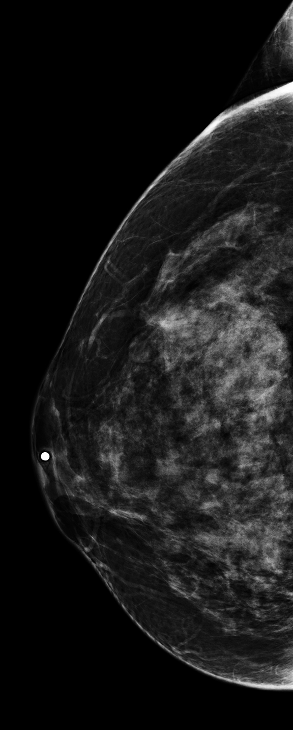
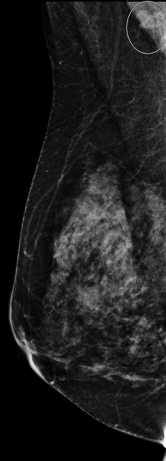
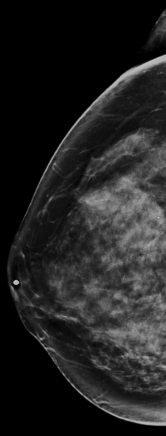
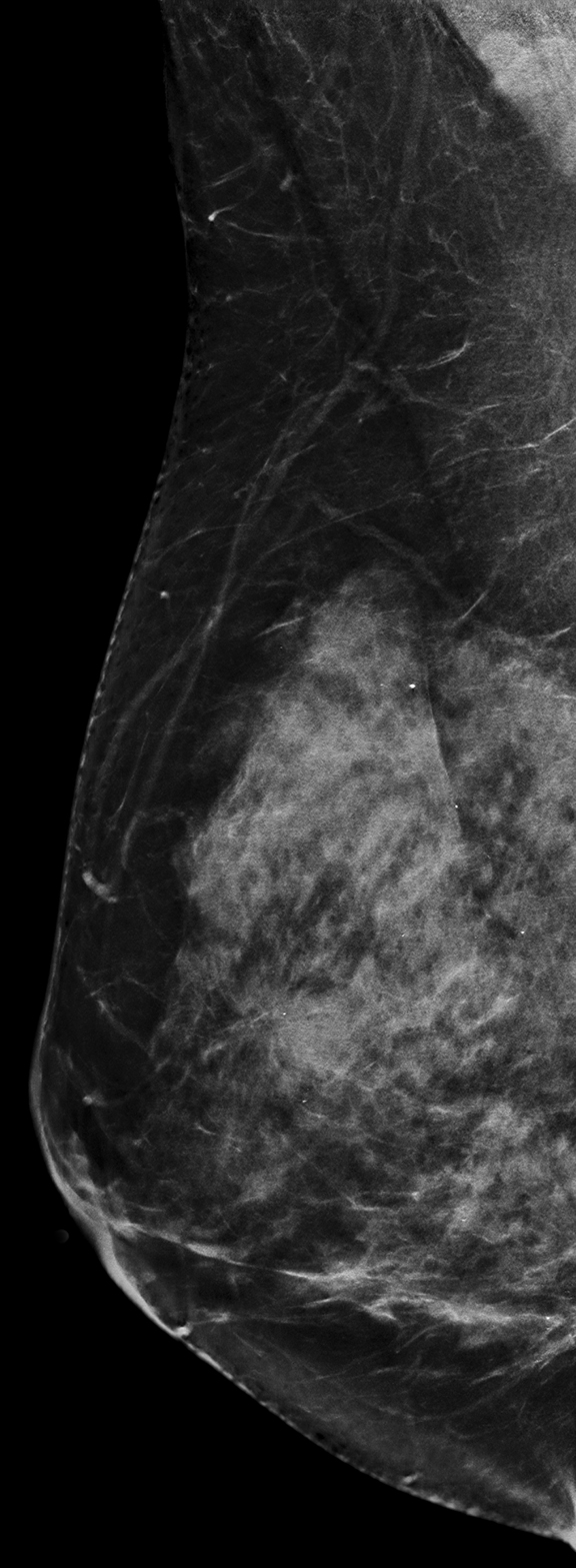
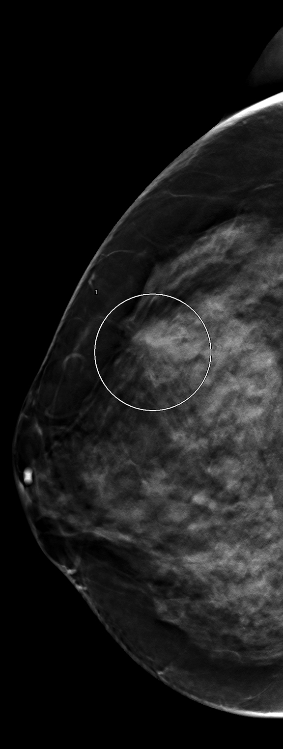
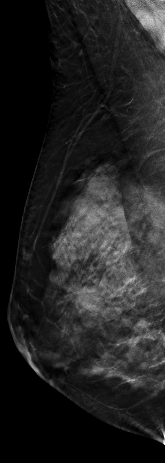
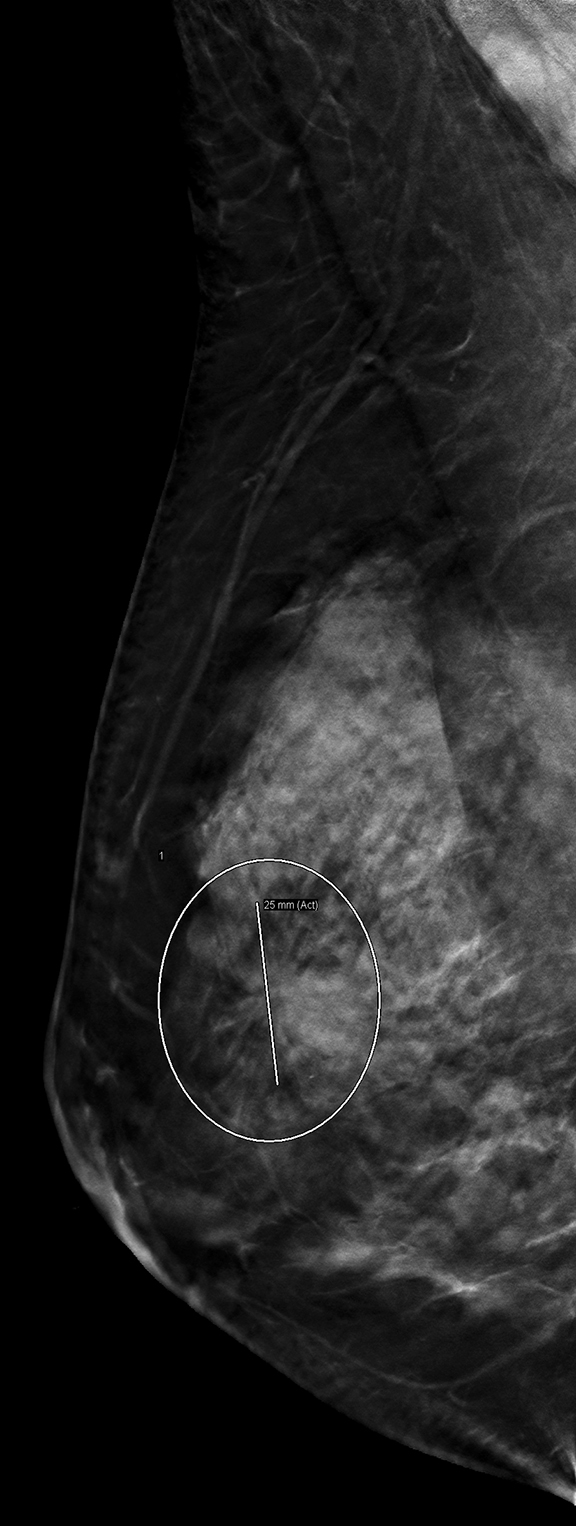
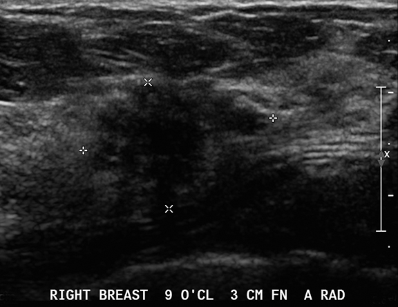
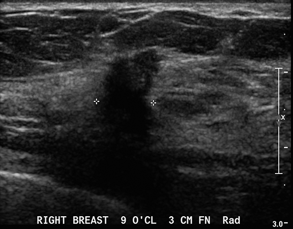
The screening mammogram was composed of full-field digital mammography (FFDM), C-view (synthesized mammogram), and digital breast tomosynthesis (DBT). There is a 2.5 cm isodense irregular mass with spiculated margins in the outer right breast. This mass is more conspicuous on C-view images (Figures 3,4) compared to FFDM (Figures 1,2) and is even more conspicuous on DBT (Figures 5-8). The diagnostic ultrasound exam (Figures 9,10) demonstrates an irregularly-shaped, hypoechoic, non-circumscribed mass with angulated margins, an echogenic halo, and posterior shadowing located 3 cm from the nipple at the 9:00 axis, which corresponds to the mammographic abnormality.
DIAGNOSIS
Invasive ductal carcinoma (IDC) with lobular carcinoma in situ (LCIS)
DISCUSSION
Digital breast tomosynthesis (DBT) is the next stage in the evolution of mammography, following on the heels of full-field digital mammography (FFDM).1-3 DBT provides multiple images at different angles as the source rotates about the breast. These images are reconstructed into 1 mm thin sections, which limit the disadvantage of overlapping dense breast tissue seen in FFDM. The combined use of FFDM and DBT (combination mode as initially mandated by the Food and Drug Administration) has led to greater diagnostic accuracy and higher positive predictive values for recall and biopsy.4-6 The addition of tomosynthesis increases the potential to identify cancers earlier and improve patient outcomes. In this case, the patient’s invasive ductal carcinoma was detected on the DBT and C-view screening mammography images before nodal metastasis occurred. Recall for spot compression was not needed.
While the combined use of DBT and FFDM decreases recall rates and detects some cancers that FFDM alone does not, the radiation dose required for the combination exam is approximately double that for FFDM alone.7 Synthesized mammography, “C-view,” is a 2-dimensional reconstruction from the DBT data set and was recently approved by the FDA. Some work has shown that it is comparable to FFDM8 and a possible substitute for the FFDM acquisition. If adopted as an alternative to FFDM, C-view addresses the concern regarding dual radiation dose.
Although great advances have been made in mammographic imaging, this modality is still limited by breast density. The patient in this case had heterogeneously dense breasts; those with heterogeneously dense or extremely dense breasts have a greater potential of having cancers obscured by overlapping normal tissue. Increased breast density results in an increased risk of breast cancer, but it unfortunately also results in a decrease in sensitivity of mammography. Ultrasound is consequently playing an expanding role as a supplementary screening modality as it has been shown to improve cancer detection.9-10
In this case, invasive ductal carcinoma with LCIS was more conspicuous on C-view compared to FFDM and even more conspicuous on tomosynthesis. Ultrasound imaging confirmed the suspicious right breast mass and allowed for minimally invasive biopsy. If neither DBT nor screening ultrasound had been performed, this cancer may have gone undetected.
CONCLUSION
Digital breast tomosynthesis detects more cancers than FFDM does. Since C-view is created from the DBT data set, it may increase the rate of cancer detection and provide greater detail compared to FFDM. This case demonstrates the usefulness of tomosynthesis and ultrasound imaging in detecting and diagnosing invasive breast cancer.
REFERENCES
- Friedewald SM, Rafferty EA, Rose SL, et al. Breast cancer screening using tomosynthesis in combination with digital mammography. JAMA. 2014;311(24):2499-2507.
- Haas BM, Kalra V, Geisel J, Raghu M, Durand M, Philpotts LE. Comparison of tomosynthesis plus digital mammography and digital mammography alone for breast cancer screening.Radiology. 2013;269(3):694-700.
- Rafferty EA, Park JM, Philpotts LE, et al. Assessing radiologist performance using combined digital mammography and breast tomosynthesis compared with digital mammography alone: Results of a multicenter, multireader trial. Radiology. 2013 Jan;266(1):104-13.
- Rafferty EA, Park JM, Philpotts LE, Poplack SP, Sumkin JH, Halpern EF, Niklason LT. Diagnostic accuracy and recall rates for digital mammography and digital mammography combined with one-view and two-view tomosynthesis: Results of an enriched reader study. AJR Am J Roentgenol. 2014202(2):273-81.
- Rose SL, Tidwell AL, Bujnoch LJ, et al. Implementation of breast tomosynthesis in a routine screening practice: an observational study. AJR Am J Roentgenol. 2013;200(6):1401-1408.
- Skaane P, Bandos AI, Gullien R, et al. Comparison of digital mammography alone and digital mammography plus tomosynthesis in a population-based screening program. Radiology. 2013;267(1):47-56.
- Samantha Zuckerman, Margolies L, Cohen S, Mandeli J, Subramaniam R, Szabo J, Patel N, Sonnenblick E. Radiation dose in the 2D and 3D components of digital breast tomosynthesis. Research. 2014;1:998.
- Zuley ML, Guo B, Catullo VJ, Chough DM, Kelly AE, Lu AH, Rathfon GY, Lee Spangler M, Sumkin JH, Wallace LP, et al. Comparison of two-dimensional synthesized mammograms versus original digital mammograms alone and in combination with tomosynthesis images. Radiology. 2014;271(3):664-671.
- Weigert J, Steenbergen S. The connecticut experiment: the role of ultrasound in the screening of women with dense breasts. Breast J. 2012;18(6):517-522.
- Hooley RJ, Scoutt LM, Philpotts LE. Breast ultrasonography: State of the art. Radiology. 2013;268(3):642-659.
Citation
E H, J S, A M, L M. Invasive breast cancer: Digital breast tomosynthesis versus full-field digital mammography. Appl Radiol. 2016;(12):32-34.
December 9, 2016