Making the Move to Digital Mammography
In recent years, digital technology has revolutionized the practice of radiology. While many clinical areas within medical imaging have already undergone a widespread migration to digital technology, mammography has been one of the last to embrace this change. Recently, however, with the publication of groundbreaking studies, such as the Digital Mammographic Imaging Screening Trial 1 (DMIST), which clearly demonstrate the benefits of this technology, many healthcare facilities have begun to make the move to digital mammography.
One healthcare organization that recently began offering digital mammography is Centegra Health Systems. Located in northeastern Illinois, Centegra is the region's leading provider of care, with access to 30 clinical sites. They perform approximately 7000 screening and diagnostic mammograms a year, averaging more than 500 such studies a month.
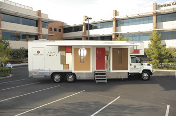
When Centegra's main campus, Northern Illinois Medical Center (McHenry, IL), needed a new stereotactic core biopsy unit, they decided to use that opportunity to make the move to digital technology. "Through the process of replacing our stereotactic core biopsy unit, we obtained a full-field digital mammography system, the MAMMOMAT Novation DR from Siemens Medical Solutions (Malvern, PA)," explained Donna Billeck-Gonshorek, RTR(M), Team Leader for the Women's Center at Northern Illinois Medical Center. This all-in-one system can be used for screening and diagnostic mammography, geometrical magnification, high-resolution spot imaging, stereotactic biopsy, specimen radiography, and galactography. It features a dual-target X-ray tube, amorphous selenium detector technology, and a large (24 × 29 cm) detector plate, allowing it to image nearly all breast sizes.
Installation and integration
Once purchased, the digital system had to be installed and integrated into the hospital's existing workflow. This process was not difficult, according to Billeck-Gonshorek. "We had some software issues at first but it was never a problem with the equipment or the detector." Most of the issues that arose, she noted, related to the integration of the new system's software with existing hospital system, such as the hospital information system (HIS), radiology information system (RIS), and the picture archiving and communication systems (PACS).
Once the system was up and running, integration with the archival system was uncomplicated as well. "Creating an archival system was not an issue for us because we had already had PACS for roughly 3 years," said Billeck-Gonshorek. "Therefore, it was just a matter of buying additional memory since digital mammograms take up an enormous amount of memory."
Digital mammography as a marketing tool
With the MAMMOMAT Novation DR in place, the organization began marketing the benefits of digital mammography to both its referring physicians and their patients. "We promote the higher detection rate in the same manner as the DMIST report," explained Billeck-Gonshorek, "as well as its value for patients between the ages of 40 and 50 years and for those with fibrocystic breasts or a history of breast cancer."
More physicians are beginning to acknowledge these benefits, she noted, and are writing prescriptions specifically for "digital mammograms." Patients are requesting digital mammograms as well, although sometimes for the wrong reason. "It seems that there is a misconception on the part of some patients that there is no compression with this technology," she said, "so patients want digital because they think it is going to be less uncomfortable. We try to explain that it is the filming that is different."
Who gets digital?
With both digital and analog mammography systems available, decisions must be made as to which technology to use for each patient. To date, Northern Illinois Medical Center has not set specific rules regarding which patients receive which type of mammogram. "If a patient requests a digital mammogram, they are given one," said Billeck-Gonshorek. "If it is not requested, those patients who are better candidates for it-those who are fibrocystic, those between the ages of 40 and 50 years, and those with a family history of breast cancer-are chosen for digital first. The remainder are chosen on a first-come-first-served basis."
Benefits of digital mammography
In the first year of using digital mammography, Billeck-Gonshorek has seen many of the benefits of this technology. "The images are incredible. I've been doing mammograms for more than 20 years and it's like night and day to me. With the digital system, we are able to visualize many more lesions. Some of the cancers that we are seeing probably wouldn't have been found at the same time with film. The patients probably would have waited another year before we caught it on an analog mammogram. That's not always the case, but that is what we are seeing in our facility. This increase in diagnostic capabilities is a benefit for the patient."
She is seeing potential benefits for the staff and institution as well. "For now, we still schedule 1 patient every half hour, but the workflow eventually will be a lot faster than it was with analog," she said. "At some point, we may be able to go to 1 patient every 15 or even every 10 minutes with digital mammograms. With digital, the pictures are instantaneous, so you are able to see right away if you need to do any extra views. If something is even questionable, you can do the spot or magnification views immediately; whereas before, you would have had to take the films, develop them in the processor, make sure they came out OK, then take more, go back, and check them again. The digital workflow is much better."
MAMMOMAT Novation DR features
Billeck-Gonshorek found several of the features specific to the MAMMOMAT Novation DR system beneficial as well. For her, one of the most important innovations was the system's unique flex compression paddle, which was designed to provide optimum levels of compression for enhanced image quality while still providing maximum comfort for the patient. The OpFocus compression plate allows for fast and easy patient positioning and keeps the breast in the central beam. It also provides for enhanced visualization of the pectoral muscle.
The OpComp feature compresses the breast only as long as the tissue is soft and pliable and automatically stops at the point of optimal compression. In addition, the SoftSpeed element slows the compression plate after its initial contact with the breast and adjusts the speed according to compression resistance.
"The Novation 's paddle is more comfortable for the patient," said Billeck-Gonshorek, "and, because it is more flexible, the paddle provides flatter, more even compression across the entire breast. In fact, our radiologists have commented on that and have said that they see a more even compression from the thick chest wall tissue to the thinner nipple tissue. It is the only paddle I use."
The system's OpDose feature automatically selects the best anode/filter combination (Mo/Mo, Mo/Rh, or W/Rh) and the lowest appropriate dose for the individual breast characteristics. This feature provides an optimized X-ray spectrum for each patient and reduces both the exposure time and dose while providing enhanced image quality. Use of the Tungsten tube (W/Rh) provides the same image quality as the Mo/Mo combination but up to 50% less dose.
With its syngo Acquisition Workstation and high-speed softcopy reporting workstation, syngo MammoReport, the Novation DR also streamlines workflow. The images are available for immediate viewing with the syngo MammoReport, which is capable of loading an 8-image case in <1 second. HIS/RIS and PACS connectivity facilitate digital archiving with the RIS client running on a separate PC and monitor.
With digital mammography the images can also be manipulated for optimal viewing. "With analog imaging, once you have a picture, that's it, it can not be changed," explained Billeck-Gonshorek. "With digital technology, however, the radiologists can window and level and manipulate the images as needed. It's phenomenal."
Lessons learned
Looking back at the process of installing and integrating digital mammography into an established analog practice, Billeck-Gonshorek offers some advice for those who wish to do the same. "Anyone purchasing a digital system should make sure that all the systems will talk to one another: PACS, HIS/RIS, and CAD," she said. "These were the only things that delayed our installation."
She also noted that the process of digitizing prior film images was more time-consuming and complex than she had anticipated. "If I knew then what I know now about digitizing previous images, I would not have done it," she said, "because with our equipment it was very involved, more than I thought it would be." Another concern she has about digitized images is the potential for lost detail. "There might be detail that is lost from the analog image when you digitize it and you just don't know," she continued. "I think it's up to each individual facility and the radiologist to decide if that is something they want."
Overall, Billeck-Gonshorek reports that at Northern Illinois Medical Center they are pleased with the advances offered by digital mammography. "The detail our radiologists can see on the digital mammograms is so much better and they are so much easier to look at than analog films," she concluded. "Now many of our radiologists don't even want to read the analog images."
- Pisano ED, Gatsonis C, Hendrick E, et al. Diagnostic performance of digital versus film mammography for breast-cancer screening. N Engl J Med . 2005;353:1773-1783.
Contact Information
Siemens Medical Solutions USA, Inc.
(888) 826-9702
www.usa.siemens.com/mammography
usa.med@siemens.com