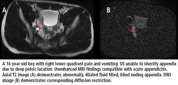
MRI exams for suspected pediatric appendicitis may not require contrast for most patients
When ultrasound findings are equivocal for pediatric patients with symptoms of acute appendicitis, radiologists are increasingly performing MRI instead of CT. The use of intravenous contrast administration for these MRI scans may not be needed for many patients, say pediatric radiologists at New York-Presbyterian Hospital/Weill Cornell Medicine in New York City.
A retrospective study published in the April issue of Pediatric Radiology suggest that accurate appendicitis diagnoses may be made without MR contrast agents for most patients. It is the protocol at New York-Presbyterian Hospital to order an MRI if an ultrasound does not show visualization of the appendix with or without suspicious secondary sonographic signs of inflammation. As a result of the study’s outcomes, Arzu Kovanlikaya, MD, professor of clinical radiology in clinical pediatrics, and colleagues have implemented a new dynamic scanning algorithm for MR investigation of pediatric appendicitis. A noncontrast scan is initially performed, with a radiologist actively monitoring the images as acquired to determine if the noncontrast images are of diagnostic quality. Intravenous contrast is administered and post-contrast imaging sequences are acquired only as deemed diagnostically needed.
The authors conducted their study to investigate the utility of intravenous contrast, and to prove or disprove their hypothesis that it might not be needed in many cases for the diagnosis of acute appendicitis. Two pediatric attending radiologists independently reviewed 112 scans of patients treated between May 2013 and January 2015. Twenty-seven patients (24%) had acute appendicitis. They conducted two reviews, one with access to the entire exam including contrast and non-contrast images, and a second time-delayed review of the exams with access to non-contrast images only. The conditional sensitivity of both groups was 100%, and the conditional specificity was 96.4% with contrast and 90.2% without contrast.
Eighty-nine scans included the use of contrast agents. In cases where the radiologists could not make a definitive diagnosis from non-contrast images alone, they were allowed access to the contrast images. In these equivocal situations, when the radiologists could add the contrast sequences, they accurately diagnosed 88 out of 89 scans compared to 78 out of 89 if only non-contrast images were available.

“In our experience, only certain cases required the use of intravenous contrast for improving diagnostic performance,” they wrote. “Only approximately one-fifth of cases required the use of post-contrast images without any commensurate decrease in diagnostic utility.Selective administration of contrast may yield similar diagnostic quality to the routine use of contrast with a substantial reduction in the number of patients receiving contrast injection.”
REFERENCE
- Lyons GR, Renjen P, Askin G. Diagnostic utility of intravenous contrast for MR imaging in pediatric appendicitis. Pediar Radiol. 2017 47;4: 398-403.