RSNA 2017 Technology: Innovation in abundance
Images
This is Part 1 of a two-part series. Part 2 will appear in the March 2018 edition of Applied Radiology.
With a call to radiologists to optimize value while embracing innovations and advancements in technology, Richard L. Ehman, MD, opened the 103rd Scientific Assembly and Annual Meeting of the Radiological Society of North America (RSNA).
In his opening address at the meeting, which unfolded Nov. 26-Dec.1 in Chicago, as it always does at the cavernous McCormick Place, Dr. Ehman, president of the RSNA, said exploring the possibilities of medical imaging and expanding its capabilities has transformed patient care.
He called radiologists “fierce early adopters of new technology” who must continue to innovate and stay focused on research. He noted that most medical imaging advances have been the result of multidisciplinary teams of physicists, radiologists, engineers and chemists working together for the rapid translation of research to clinical practice.
While artificial intelligence and machine learning were hot topics at RSNA 2017 (and more to come on these technologies in the March 2018 issue Applied Radiology), MR, breast and CT imaging were the dominant stars of 2017 scientific papers. Dose reduction remained on the forefront of CT imaging innovations and is now also being widely discussed by many (if not all) of the digital radiography vendors. For more news on clinical studies presented at RSNA 2017, visit appliedradiology.com and search for RSNA 2017.
Meanwhile, in the twin exhibit halls, a multitude of new technologies was being shown off at the most recent edition of radiology’s biggest show of the year. Here we present a modality-by-modality sampling of some of it:
Breast imaging
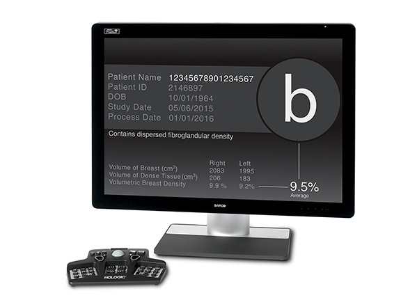
Hologic, Inc. announced U.S. FDA 510(k) clearance for its Quantra 2.2 Breast Density Assessment Software, which enables clinicians to provide women with consistent breast density assessments during routine breast cancer screenings. Through a proprietary algorithm powered by machine learning, Quantra software analyzes mammography images for distribution and texture of breast tissue, delivering clinicians patient-specific breast density assessment. Quantra software categorizes breasts into four categories of density, in alignment with the ACR BI-RADS Atlas 5th Edition.
Quantra software is available for use with Hologic 3D Mammography systems, including the new 3Dimensions mammography system, pending U.S. FDA 510(k) clearance and not commercially available, with the Intelligent 2D imaging technology. The 3Dimensions system offers a variety of features designed to provide higher quality 3D images for radiologists, enhanced workflow for technologists, and a more comfortable mammography experience, with low-dose options, for patients.
Transpara from ScreenPoint Medical is a machine learning software application designed to assist radiologists with the reading of conventional mammograms and digital breast tomosynthesis exams. The software automatically identifies soft-tissue and calcification lesions and combines the findings of all available views into a single cancer suspiciousness score using state of the art image analysis and deep learning technology. This interactive decision support system categorizes every mammogram on a 10-point scale based on calcification and soft tissue lesion findings. Intelligent software correlates MLO and CC views when abnormalities are visible in both views. Using the Transpara Score, mammograms can be sorted according to the likelihood that breast cancer is present and detectable. If no potential abnormalities are found, a low score is assigned and when there are suspicious findings, a higher score is assigned. Transpara can be used to automatically pre-screen and identify with high confidence patients who need increased attention as well as patients for whom no signs of cancer are present.
Siemens Healthineers has unveiled its new premium mammography platform, the MAMMOMAT Revelation, awaiting U.S. FDA 510(k) clearance. A new integrated specimen imaging tool facilitates immediate control of the biopsy directly at the mammography system. The MAMMOMAT Revelation also provides automated breast density measurements at the point of care. It is also designed to expand existing diagnostic options and provides patient access to functional breast imaging with contrast-enhanced mammography. The MAMMOMAT Revelation has 50-degree wide-angle 3D HD breast tomosynthesis, which offers the widest scan angle available, according to Siemens. The scan angle is the basis for the high depth resolution, which provides extremely high-quality 3D images to increase diagnostic confidence and enable earlier detection of even subtle lesions. With MAMMOMAT Revelation, the clinician also can perform biopsies based on 3D HD Breast Tomosynthesis. The HD Breast Biopsy solution enables one-click targeting of suspicious areas with a +/- 1 mm accuracy.
GE Healthcare’s newest mammography system, Senograph Pristina, incorporates the Senographe Pristina Dueta, a patient-assisted wireless remote control mammography device that enables women to manage their own compression. After the breast is properly positioned by a technologist, the patient has the option to adjust her compression. The device does not compromise image quality or exam time.
The next generation of SenoBright HD contrast enhanced spectral mammography (CESM) is also being demonstrated. It is used with Senograph Prestina. Compared to the first generation of CESM technology, SenoBright HD delivers clearer images and improves acquisition time by 40 percent in women with large breasts, according to GE Healthcare. Images are immediately available for review following an exam.
The ASPIRE Cristalle with DBT option from FUJIFILM Medical Systems U.S.A., Inc., combines state-of-the-art, Hexagonal Close Pattern (HCP) image capture technology and intelligent image processing to help optimize contrast and dose based on auto recognition of breast characteristics. Its patented, flexible Comfort Paddle provides noticeable patient comfort, even with compression of the breast. The company also offers its Dynamic Visualization II for Mammography (DVIIm) image processing software with the ASPIRE Cristalle (FFDM). The second-generation software now offers intelligent auto recognition of breast tissue, implant and structural characteristics to intelligently adapt image contrast and density, for enhanced diagnostic visibility. DVIIm provides improved contrast and density stability throughout the entire exposure region and achieves improved visibility across a wide range of breast compositions including the presence of implants.
Ikonopedia introduced new breast reporting and analytics tools to support compliance with the new U.S. FDA-mandated image quality requirements of the Enhancing Quality Using the Inspection Program (EQUIP). Ikonopedia automatically initiates periodic U.S. FDA-mandated image quality reviews, for both interpreting physicians and radiology technicians, to review a sample set of auto-generated exams, document deficiencies and indicate required corrective actions. Each exam is graded on the eight standard image quality measures, and grading criteria can be customized. Ongoing oversight of QA/QC records and corrective action is managed through Ikonopedia Analytics, with reports specifically designed to address EQUIP compliance. For mobile clinics, multiple mobile stops can be grouped together as a single virtual site for quality review purposes. Also new, the Patient Canvas is part of Ikonopedia’s Patient View module, a secure overview of all pertinent information about a patient. Patient Canvas is a graphical drag-and-drop scar/mole/lesion locator used by technologists, which can be made available to radiologists as they are reading images.
Ikonopedia and Konica Minolta Healthcare Americas, Inc. announced they have expanded their partnership to integrate both the Opal and Exa healthcare IT platforms from Konica Minolta with Ikonopedia’s solution for breast imaging reporting and tracking. The integration with Exa offers a seamless, closed-loop experience with multi-directional functionality and automated follow-up for patients with significant BI RADs. Ikonopedia’s solution captures various points of data from the Exa Mammo software to provide a complete patient profile that will result in automatic patient warnings and alerts.
By leveraging artificial intelligence (AI), PowerLook Tomo Detection from iCAD, Inc. can optimize digital breast tomosynthesis (DBT) reading efficiency and improve clinical confidence. This efficiency enables users to streamline workflow and supports faster, more confident detection of breast cancer. According to the company, PowerLook Tomo Detection is the first and only U.S. FDA cleared concurrent-read cancer detection solution for DBT.
PowerLook Tomo Detection utilizes a trained algorithm developed through deep learning that automatically analyzes each tomosynthesis plane. Suspicious areas identified are then blended into a 2D synthetic image to provide radiologists with a single, highly sensitive, enhanced image that is used to easily navigate the tomosynthesis datasets. While PowerLook Tomo Detection is currently only available for GE Healthcare’s DBT platforms, iCAD is developing a multi-vendor solution that is expected to be available in 2018.
Seno Medical Instruments, Inc. announced positive data from PIONEER, a Phase III pivotal trial of its Imagio breast imaging system that uses opto-acoustic ultrasound (OA/US) imaging to differentiate benign from malignant masses. The study found that OA/US was more specific than device gray-scale ultrasound (US) alone in differentiating malignant from benign breast lesions and was non-inferior to US with respect to sensitivity. PIONEER was a U.S., prospective, multi-institutional study that enrolled 2,105 women over the age of 18 years.
Several key findings from the study include: In the intent to diagnose (ITD) population of 1,739 subjects and 1,808 masses, diagnostic specificity for benign masses was 43 percent for OA/US and 28.1 percent for US, corresponding to a 14.9 percent absolute specificity advantage for OA/US over US (p<0.0001); for masses that were benign, OA/US resulted in downgrading 34.5 percent of US reads (from BR4A to ≤BR3 or BR3 to BR2). OA/US also resulted in upgrading 6.0 percent of US mass reads, for a net downgrade rate of 28.5 percent (≥0.0001); and for masses that were malignant, OA/US resulted in upgrading 47.0 percent of mass reads classified as BR3 by US and downgrading to BR2 27.3 percent of mass reads classified as BR3 by US. Positive predictive value for masses assessed as BR4A or higher was 51.5 percent for OA/US compared with 46.3 percent for US; negative predictive value for masses assessed as BR3 or lower is 94.4 percent for OA/US compared with 97.0 percent for US.
Computed tomography
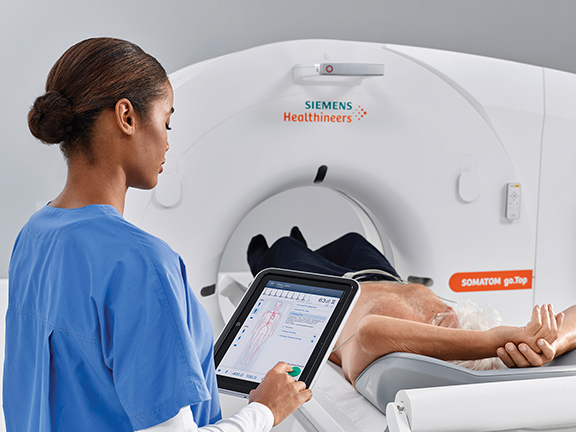
Siemens Healthineers expanded its tablet-based CT workflow platform, SOMATOM.go, into cardiology and CT-guided interventions with the introduction of SOMATOM.go.All and the SOMATOM go.Top. The scanners are operated via a tablet that can be used to control all routine and advanced examinations. The 128-slice SOMATOM.go.Top can perform whole-body scans of up to 200 cm with a scan speed of up to 175 mm per second, and the 64-slice SOMATOM.go.All can cover scan ranges of up to 100 mm in one second. A new X-ray tube allows users to adjust the tube voltage in 10-kilovolt increments while keeping the tube current high.
The company also introduced SOMATOM Edge Plus, a new premium, single-source scanner, and the SOMATOM Force, a new version of a high-end dual source scanner equipped with two X-ray tubes and two detectors. The new systems allow clinical users to cover all CT applications, regardless of patient or clinical issue. Siemens states that the systems’ precise diagnostics come from the FAST applications integrated into the technologies. One such application is the FAST Integrated Workflow with a new FAST 3D Camera for automatic patient positioning. Currently pending U.S. FDA 510(k) clearance, this innovation is designed to help users eliminate position misalignment.
The latest configuration of the spectral CT from Royal Philips provides clinicians with advanced functionality that supports emergency and trauma department care. The new IQon Elite CT has faster reconstruction speeds and better visualization of bone marrow pathology. These faster reconstruction speeds have been shown to enable the imaging of up to 200 CT patients per day (under a protocol of 600 images per SBI, 3 conventional series per patient and 1.5 SBI). The scanner’s ability to estimate electron density enhances tissue characterization, while a new radiation therapy planning couch and bariatric table permit larger patients to be scanned with increased positioning controls. The IQon Elite Spectral CT scanner will be available globally in the first quarter of 2018.
The Aquilion Precision from Toshiba Medical, a Canon Group Company is an ultra-high resolution CT scanner capable of resolving anatomy as small as 150 microns that can provide CT images with resolution typically seen only in cath labs. Toshiba Medical says the newly designed detector provides more than twice the resolution of current technology. The system has a focal spot tube at 0.4 mm x 0.5 mm and a 1024 and 2048 reconstruction matrix. Features to improve dose efficiency include detector channels that are 0.25 mm thick, as well as substantial improvements in scintillator quantum efficiency, detector circuitry and other DAS components. The Aquilion Precision is pending U.S. FDA 510(k) clearance.
Also new, the Aquilion Prime SP can generate 160 unique slices per rotation with 0.35-second scanning capability. It features a 78-cm aperture gantry and a 660-pound patient weight-capacity couch. An optimized beam spectrum and a more efficient detector based onPUREVISION Optics provides the Aquilion Prime SP with reduced dose by up to 31 percent and improved LCD for the body (22 percent) and brain (19 percent).
Revolution Frontier represents the latest addition to GE Healthcare’s CT product portfolio. The system includes an entirely new imaging chain, the Gemstone Clarity detector and Performix HD Plus X-ray tube featuring liquid bearing X-ray tube technology that the company says delivers 2x longer life. The system enables visualization of fine anatomic detail of 0.23 mm with 25 percent lower electronic noise. When used with Revolution Frontier, GSI Pro creates 2x faster reconstruction of spectral images. Scans using GSI Pro can deliver tissue characterization, contrast dose reduction, metal artifact reduction and quantitative information about chemical composition. Plus, ASiR-V enables dose-neutral GSI and routine HD anatomic detail.
GE also announced Smart Subscription, an offering that provides continuous access to the latest software and applications for a hospital or clinic’s CT devices, for one fee per device per year. Smart Subscription will help: avoid obsolescence throughout the lifecycle of a system; ensure the same CT capabilities at all sites provide consistent exams throughout a healthcare enterprise; increase staff efficiency, reduce training and improve satisfaction by ensuring there is one set of capabilities to learn, operate and read.
Debuting at RSNA was Samsung Electronics’ OmniTom® mobile, 16-slice computed tomography (CT) scanner. OmniTom features omni-directional wheels, maximizing mobility and allowing easier and quieter movement in small spaces. The 16-slice (0.625 mm per slice) advanced data acquisition system delivers a 50-percent increase in CNR versus other portable CT technologies. A small footprint is ideal for mobile use yet it has a 40-cm gantry opening for improved coverage of adult head and neck, and full-body pediatric scanning. OmniTom features an internal drive system, making portability less strenuous, while also offering smart-sensing collision avoidance software to maximize control and patient safety.
Carestream Health demonstrated new optional advanced metal artifact reduction software for its CARESTREAM OnSight 3D Extremity System pending. The software, pending U.S. FDA 510(k) clearance can be adjusted and optimized according to the amount of metal present. It uses information from the original scan to eliminate additional imaging studies. An intuitive touch screen interface allows technologists to adjust for either moderate or complex metal content. The metal artifact reduction software can be activated prior to the scan or it can be applied after the original reconstruction is complete. Both the original and corrected images are always available to view and compare.
Digital radiography
Konica Minolta Healthcare Americas, Inc. made several new product announcements for its line of DR solutions. REALISM, an advanced image processing solution, delivers a new level of clarity and detail for superior visualization within soft tissue and bony structures. By independently processing bone and soft tissue data, it enhances X-ray image sharpness and contrast to reveal subtle aspects of the image, even in the most difficult anatomies. Along with improvements in image quality, REALISM can enhance workflow efficiency by enabling visualization of soft tissue and bone simultaneously, reducing the number of window-level adjustments needed.
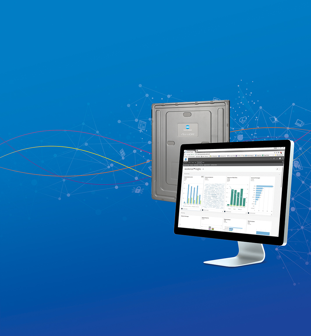
The company launched two additional sizes of its AeroDR HD wireless flat panel detectors. The new 10” x 12” AeroDR HD detector is ideal for imaging fine structures such as extremities and for the special needs of NICU environments, and the new 17” x 17” AeroDR HD is designed for imaging larger anatomical areas, like the chest and abdomen. All AeroDR HD panels offer the option to switch between High Definition (HD) and High Dynamic Range (HDR) imaging.
Konica Minolta Healthcare also demonstrated new software, AeroRemote Insights, a web-based tool designed for use with the company’s DR systems. AeroRemote Insights delivers valuable, interactive and actionable data about productivity, user performance and imaging efficiency and effectiveness. Managers can remotely evaluate productivity by technologist or exam room and identify areas for improvement, such as exam repeat rates. The software also assesses the health of the Konica Minolta AeroDR systems in use to ensure optimal calibration and performance. The new software will be available for most Konica Minolta AeroDR systems in early 2018.
Canon U.S.A. and its wholly owned subsidiary Virtual Imaging exhibited the newly released CDI-710C, CXDI-810C and CXDI-410-C Wireless Detectors. The detectors are lightweight and IPX7 rated devices that feature limited on-board image storage capability when operated in Standalone Mode. The company states that by using strong carbon fiber, the new Canon detectors are among the lightest weight detectors currently available and are designed with form and function in mind. A carbon fiber chassis and frame provide high performance and high durability. Each of these three wireless detectors features a “ready” function to be used when working with multiple detectors in a single environment. This function enables a user to select a specific detector from the DR modality workstation by simply pressing the “Ready” button.
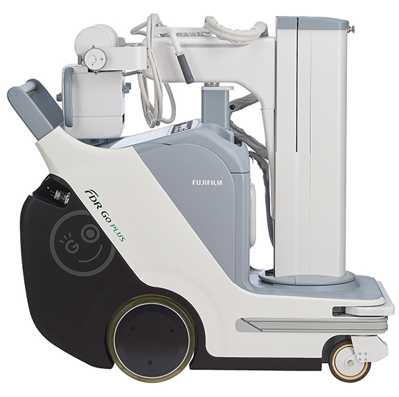
The all-new FDR Go PLUSversion portable digital radiography system was on display in the FUJIFILM Medical Systems U.S.A., Inc. booth. In addition to featuring a redesign, its smooth, quiet travel and compact tube head, the system now includes a collapsible column for maximum visibility while traveling. The portable DR system also features an extra-large display for previewing images at the bedside. Other enhancements include user adjustable drive handle (option), wireless barcode reader (option), an RFID card reader (option) and extensive dedicated storage areas. Software features of the new portable DR system include new workstation software with automated keypad display, quick start, the company’s latest Virtual Grid simulation software (option) and Dynamic Visualization II image processing (option).
FUJIFILM and FUJIFILM SonoSite also introduced a comprehensive pediatric imaging solutions portfolio consisting of digital radiography systems, point-of-care ultrasound system, and pediatric-focused healthcare IT solutions. The pediatric portfolio is designed to combat challenges associated with pediatric imaging, such as minimizing dose exposure, increasing speed of image acquisition, helping improve infection controls, and reducing patient anxiety associated with exams. DR products in the pediatric portfolio include the FDR AQRO, FDR D-EVO II CsI, FDR D-EVO GL and FDR Go PLUSversion Digital X-ray System.
Carestream Health showcased its new CARESTREAM DRX-Revolution Nano Mobile X-ray System that uses carbon nanotube technology to deliver significantly reduced size and weight when compared to existing mobile X-ray systems. The compact, 200-pound, non-motorized system is easier to move and position even in cramped critical care areas and includes a fully integrated digital workflow. Additional features include an advanced lithium iron phosphate battery that, along with the carbon nanotube technology, contributes to longer life and a sleek design with enhanced visibility both over and around the system. The CARESTREAM DRX-Revolution Nano Mobile X-ray System can be used with all Carestream DRX detectors and it is scheduled for availability in 2018.
The new Discovery XR656 HD premium fixed digital radiography system from GE Healthcare features an expanded suite of workflow automation and analytics tools and includes the new Helix advanced image processing platform. It is designed to eliminate unnecessary X-ray image adjustments and repeat exams. When using FlashPad HD digital detectors, images with up to 4x higher definition in each image may be acquired. The FlashPad HD digital detectors improve contrast detectability by up to 40 percent due to ultra-high resolution, enhanced noise control, and advanced image processing. The Helix advanced image processing is being touted by GE Healthcare as a revolutionary platform that features advanced image processing, ultra-high resolution digital detectors, powerful analytics, and intelligent systems. Helix delivers sharp detail at low dose and consistent performance in radiography, despite variations in exposure technique and patient anatomy.
GE also introduced its newest mobile X-ray system, the Optima XR240amx with FlashPad HD digital detectors that enable clinicians to see very fine detail in all anatomies with its acquisition of high-resolution images. Highly maneuverable and compact, it offers seamless X-ray imaging support in neonatal environments. Two detectors can be charged simultaneously in-bin, with or without grids attached. The system can support imaging 50 patients over 9.5 hours, according to the company.
Agfa HealthCare demonstrated its multipurpose DR 800 X-ray room, a work in process, enriched with Dynamic MUSICA image processing and the new X-Team Technology for viewing and communicating. The integrated solution can handle a full range of radiography (including static exams and tilting exams) and fluoroscopy exams (including barium studies, arthrograms, cystograms, myelography and catheter placement, etc.). The DR 800 comes with Dynamic MUSICA high-speed, multi-scale image processing, which can now also process moving images, such as fluoroscopy. Dynamic MUSICA also enhances noise suppression, stabilizes brightness, reduces veiling glare, and can play a significant role in enabling potential dose reduction.
Agfa HealthCare also showcased its new X-Team Technology. This workflow enrichment integrates the XERO universal viewer, which provides secure access to images from different departments and sources, in one view, in the MUSICA workstation. Collaboration features, such as instant messaging and clinical dialogue, enhance consultation on study acquisition between radiologists and radiographers.
The S-Vue engine in Samsung’s Ceiling Digital X-ray GC85A recently received U.S. FDA clearance. S-Vue provides reliably clear images, enhanced sharpness and clarity, and adjustable contrast presets. A precise auto shutter improves consistency. Unique to the GC85A is the spatially adaptive multi-scale processing and advanced de-noising technology that enable high quality images even at half the dose.
Magnetic resonance imaging
The SIGNA Premier wide bore 3.0T MRI system, which received 510(k) clearance from the U.S. FDA in August 2017, features GE Healthcare’s latest, short-bore, high-homogeneity 3.0T superconductive magnet, the most powerful gradient system the company has ever developed for a wide bore 3.0T system, and new, digital RF transmit and receive architecture. It enables two times faster whole-body imaging compared to conventional MRI scans, and 60 percent faster images. SIGNA Premier is a result of a four-year collaboration with the National Football League and research institutions around the world to aid researchers in the detection of biomarkers for the potential diagnosis of mild traumatic brain injury
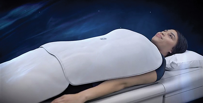
A new suite of RF coils, AIR Technology, is now commercially available on SIGNA Premier. AIR Technology uses a flexible conductor material that allows each coil element to be closer to the anatomy to improve signal reception, depth of penetration and image quality. The ultra-lightweight design of the coils also makes it easier for a technologist to position a patient. The coils are 60 percent lighter on the patient, have the appearance of a blanket and incorporate technology engineered for higher density channel count and increased signal-to-noise ratio (SNR).
Hitachi Healthcare Americas demonstrated its Synergy Drive MR workflow engine, which is now standard with Hitachi’s Echelon Oval 1.5T MR system. SynergyDrive addresses speed of throughput with automated patient registration, pre-population of scanning protocols, streamlined patient positioning and setup. Its Auto Image Load feature helps efficiently prepare the next scan for seamless transition. SynergyDrive also includes a number of sequences and image processing tools to improve image quality and reduce patient callbacks. RADAR motion compensation, FatSep, RAPID parallel imaging and non-contrast MR angiography image sequences deliver fast, high quality images while reducing the amount of time a patient must remain motionless.
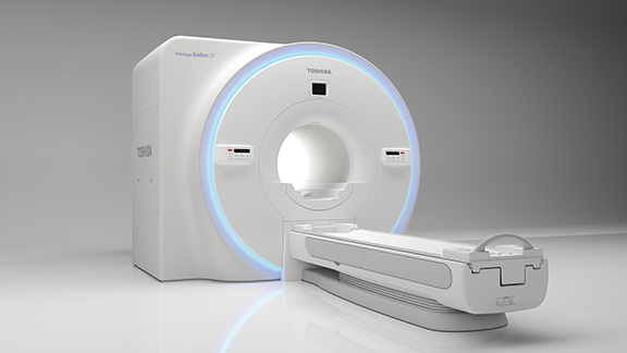
Two new MRI scanners were introduced by Toshiba Medical. The company rolled out Vantage Galan 3T, designed to enhance oncology radiation therapy by allowing healthcare providers to precisely map MR data to PET/CT images. It includes the Universal Couchtop MR Overlay from CIVCO Radiotherapy to aid clinicians in accurately determining the treatment target area, a Three-Pin Lok-Bar offers providers the option to use existing MR-compatible devices by indexing them to the overlay, and adjustable ATLAS body coil technology that allows clinicians to combine coils for extended patient coverage and enhanced workflow.
The Vantage Elan/Zen Edition 1.5T has an easy-to-use interface designed to improve workflow and productivity. Automatic slice alignment with EasyTech standardizes workflow with automatic positioning while delivering consistent image quality. Ultrashort Echo Time (UTE) captures images in tissues that generally disappear too quickly for accurate MR imaging, enabling imaging of anatomy such as the MSK and lungs. Pianissimo Zen reduces sound during acquisition by 99 percent. For cardiac imaging, multi-echo T2 Mapping maps with Toshiba Medical’s updated FFE2D mEcho sequence can be used in quantification and analysis of myocardial iron overload. T1 mapping that utilizes MOdified Look-Locker Inversion recovery (MOLLI) sequence allows providers to acquire a more quantitative characterization of myocardial tissue within a single breath hold.
While Siemens Healthineers showcased its newest 3T MRI scanner, MAGNETOM Vida, some of the biggest pre-RSNA MRI news was the U.S. FDA’s clearance of the company’s MAGNETOM Terra 7T MRI. The Terra 7T delivers up to 64 receive channels and 2x the SNR of 3T MRI in optimized 7T neuro and MSK clinical applications. The 7T scanner provides ultrafine 0.2 mm in-plane anatomical resolution. Its 80/200 gradients provide the power needed to perform diffusion MRI and functional MRI (fMRI), and enables use of the company’s Simultaneous Multi-Slice application to accelerate advanced neurological applications for clinical routine.
MAGNETOM Vida features new BioMatrix technology that addresses anatomical and physiological differences among patients, as well as user variability. BioMatrix Sensors built into the scanner’s new patient table automatically track respiratory patterns as soon as the patient lies on the table. BioMatrix Tuners can improve the quality and reproducibility of whole-spine diffusion imaging via individual slice adjustments that mitigate distortion that otherwise may occur, especially at 3T. Biomatrix Interfaces help ensure consistently high exam quality, accelerating scanning by up to 30 percent and improving patient care.
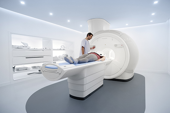
MR Prodiva 1.5T is the newest MR system from Royal Philips that provides enhanced clinical performance, workflow and patient experience. Breeze Workflow provides a simplified, guided patient setup and includes a flexible lightweight digital coil system to support fast patient setup. The system also features Philips’ Ambient Experience In-bore Connect, which allows patients to personalize their environment with a visual theme, guiding them through the examination with instructions, and by reducing acoustic noise. Philips also announced Compressed SENSE, an application that enhances productivity in imaging by increasing the data that is able to be pulled quickly from scans, including both 2D and 3D scans, all anatomical contrasts and all anatomies. 3D APT rounds out the company’s new MR technologies; this contrast-free imaging solution supports neuro-oncology clinicians. MR Provida 1.5T, Compressed SENSE and 3D APT are all pending U.S. FDA clearance.
Perspectum Diagnostics Ltd, announced U.S. FDA 510(k) clearance for LiverMultiScan, a post-processing software device for MR imaging of the liver, delivered through a cloud-based service. This latest clearance means that this unique technology can now be used on a wider range of scanners, including compatible Siemens and Philips MR systems, and provide clinicians with standardized, quantitative measures of the liver tissue to assist with diagnosis of liver disease. LiverMultiScan enables non-invasive and quantitative liver tissue characterization to quickly and accurately quantify liver fat, as well as T2* and iron-corrected T1 which correlate to iron and fibro-inflammatory levels, respectively. It is a rapid and scalable technology that can be seamlessly integrated into existing MR examinations, without the need for contrast agents.
Ultrasound
FUJIFILM Medical Systems U.S.A., Inc. and FUJIFILM SonoSite introduced a comprehensive pediatric imaging solutions portfolio consisting of digital radiography systems, point-of-care ultrasound system, and pediatric-focused healthcare IT solutions. The pediatric portfolio is designed to combat challenges associated with pediatric imaging, such as minimizing dose exposure, increasing speed of image acquisition, helping improve infection controls, and reducing patient anxiety associated with exams. The ultrasound products in the pediatric portfolio include SonoSite X-Porte, a point-of care ultrasound system with the XDI (Extreme Definition Imaging) proprietary beam forming technology and VevoMD, an ultra-high frequency ultrasound system with transducers designed for infants and small patients to help visualize tiny anatomy not visible with conventional ultrasound.
A new work-in-progress for the Aplio i-series ultrasound family of products from Toshiba Medical is an entry-level system offering enhanced workflow and performance features. The system uses the same transducers as the current Aplio Platinum Series. It provides intuitive ergonomics to help boost productivity during daily routine and complex examinations with iSense. An image-guided user interface visually guides a clinician through the examination to simplify system operation and help improve efficiency. The iPerformance features deliver advanced imaging capabilities and applications that also help health care providers make more confident diagnoses. The Aplio i600 is currently pending 510(k) clearance from the U.S. FDA.
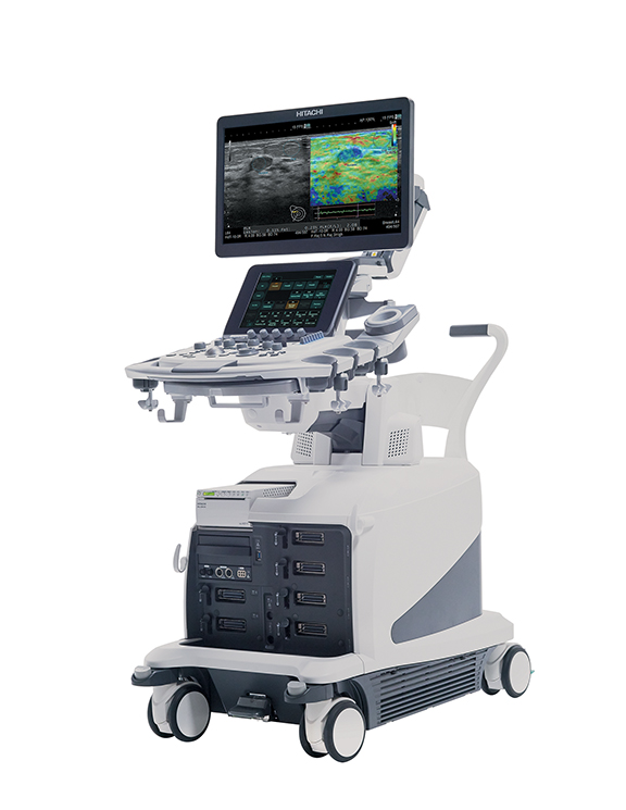
Hitachi Healthcare Americas introduced a new premium ultrasound system, the ARIETTA 850. The system builds upon the advanced capabilities of Hitachi’s ARIETTA family of ultrasound scanners, adding features like eFocusing, which removes the need for focal zone adjustments by dynamically focusing from the near to far-field of the image. It also expands interventional capabilities through a collection of features that work in conjunction with Hitachi’s Real-time Virtual Sonography fusion software to automatically perform fusion registration, compensate for needle flexion during biopsy procedures, and deliver real-time visual maps of estimated RF ablation zones. ARIETTA 850 also supports the SML44 ultrasound probe that uses capacitive micro-machined ultrasound transducers (CMUT) rather than piezoelectric crystals to transmit and receive the ultrasound signal. This design enables an ultra-wide bandwidth of 2-22MHz, allowing a single probe to address multiple clinical needs and to adapt to a wide variety of body habitus.
CIVCO Medical Solutions showcased its latest guidance technology, the Verza guidance system, designed to simplify ultrasound-guided interventions. A key feature of the Verza system is its direct transducer attachment in combination with VerzaLink, a proprietary locating feature designed onto the ultrasound transducer, eliminating the need for a custom bracket attachment and providing an ideal profile and position on the ultrasound probe. The Verza system was developed to meet the needs of clinicians accustomed to fixed-angle guidance as well as those who prefer a free-hand technique. Offering a selection of five potential approach angles, the Verza guide provides an optimal pathway for a wide assortment of anatomical targets. With 14 instrument inserts, ranging in size from 25 gauge to 12 French, Verza is designed for compatibility with the market’s largest range of interventional devices. Verza is now available for GE Healthcare LOGIQ E9 XDclear 2.0, Philips EPIQ with Evolution 4.0, and Toshiba iAplio iSeries i700, i800, and i900 systems.
Other technology
Contrast media
Guerbet launched Contrast&Care, a new contrast media injection management solution that recently received CE Mark approval. Contrast&Care is an integrated information technology solution that groups together all the information related to exams requiring the injection of contrast media (contrast media, injection protocol, patient data, etc.). It seamlessly interfaces with information systems, such as RIS and PACS. The information generated by compatible injectors is transmitted automatically to Contrast&Care at the end of each exam. This enables users to view the history of each patient, the exams with injection that have been performed and the administered doses of contrast media. Contrast&Care also offers the option of reviewing protocols, creating protocol libraries and viewing statistics and trends relative to injection activity and the use of contrast media.
On the heels of its recent U.S. FDA approval, TAGITOL V (barium sulfate) oral suspension from Bracco Diagnostics was available for the first time at RSNA. The low-volume fecal tagging agent is used to opacify residual stool in the colon for CT imaging. According to Bracco, TAGITOL V is the first contrast imaging agent approved by the FDA for tagging residual material during a CT colonography (CTC), also known as virtual colonoscopy, which helps clinicians differentiate true polyps from stool and could lower false positives.
The company also announced its development and sponsorship of www.myctcolonography.com, a website dedicated to educating patients and their primary care physicians about CTC testing.
Citation
CE K, M M. RSNA 2017 Technology: Innovation in abundance. Appl Radiol. 2018;(1):20-29.
January 11, 2018