Updates in breast imaging: TMIST, guidelines and tomosynthesis technology
Images
It’s been a decade since the pivotal DMIST study determined that digital mammography delivers greater breast cancer detection accuracy than film-screen mammography in women under age 50 and those with radiographically dense breasts. However, since then there has been a growing understanding of the limitations of digital mammography screening in women with dense breasts, which can often make it difficult to detect lesions. Digital breast tomosynthesis is one possible answer to this dilemma.
“Tomosynthesis goes one step further than digital mammography,” says Martin Yaffe, PhD, CM, Senior Scientist at Sunnybrook Health Sciences Centre in Toronto. Dr. Yaffe explains that tomosynthesis images present the perspective of looking through the breast, eliminating the superposition of shadows from the different layers of breast tissue that can mask cancer or create patterns that resemble cancer when none is present.
The potential benefits of tomosynthesis include finding cancers not detected by digital mammography, reducing false positives, and more precisely locating and sizing the lesion(s) for better assessment of the disease. Dr. Yaffe cautions against calling tomosynthesis “3D,” explaining that it creates images that look three dimensional, but the images are produced differently from, for example, 3D CT images. Yet, he believes tomosynthesis does deliver important technological advantages over digital mammography.
“The biggest impact will be the improvement in sensitivity and specificity in screening,” adds Dr. Yaffe, who has been working with Etta Pisano, MD, Vice Chair of Research and staff radiologist, breast imaging, at Beth Israel Deaconess Medical Center (Boston) on a trial aimed at evaluating tomosynthesis for breast cancer screening. The Tomosynthesis Mammographic Imaging Screening Trial (TMIST) will be a multi-center, randomized trial comparing the diagnostic accuracy of tomosynthesis against 2D full field digital mammography (FFDM), the current standard of care for breast screening.
Dr. Pisano, who is also on the faculty of Harvard Medical School, said TMIST was conceived five years ago as a traditional technology assessment. However, it has evolved into a true breast screening trial. The last such trial, she says, was over 30 years ago. In the early 1980s, The Health Insurance Plan [HIP] of Greater New York was the first published large-scale clinical trial that demonstrated the efficacy of mammography.1 It was followed by the Two-County study in Sweden.
While technology and therapy have improved over the last few decades, screening is still being questioned as a necessary annual exam, particularly for women under 50.
“TMIST is a way to prove that what we find (with breast screenings) are the most important cancers,” Dr. Pisano says. “I believe this study will show that this newer technology reduces advanced breast cancers in the population diagnosed with the disease.”
TMIST will examine the efficacy of the technology, and that is a fundamental shift in the way radiology conducts research, Dr. Pisano says. The study is designed to provide direct evidence of the benefits of screening, specifically with tomosynthesis.
Women will be randomized for screening with DBT or FFDM and then followed for four years. “We think tomosynthesis will find the cancers earlier and women with dense breasts are more likely to have an advantage of earlier diagnosis with it,” Dr. Pisano adds.
Supported by a grant from the Canadian Breast Cancer Foundation, Dr. Yaffe has already begun the Canadian arm of the TMIST trial in Toronto, Vancouver and Ottawa. Since first recruiting patients in November 2014, the Canadian study has enrolled 2,000 women.
Altogether, TMIST will enroll 150,000 women in 90 centers throughout Canada and the U.S. Dr. Pisano is confident the National Institutes of Health will help fund TMIST in the U.S., and she hopes that funding will be secured by the end of 2016.
Expectations are high for the TMIST study. Based on previously published smaller or single-site studies, Dr. Yaffe hopes to see a reduction in recall rates of 30-35% with women who have false positives, based on pathology.
“With tomosynthesis, we have clearer images, so we expect that to lead to fewer false positives. The key is to find the lesions that are likely to grow more aggressively,” he says. “We are looking at whether, by catching them earlier, we can reduce the future incidence of the more life threatening breast cancers, those that are node positive or that have more aggressive molecular characteristics.”
Dr. Yaffe says tomosynthesis is not reimbursed nor widely adopted as a screening tool in Canada. In the U.S., where tomosynthesis is not reimbursed by most private payers, but is by the Centers for Medicare & Medicaid Services, Dr. Pisano says the latest FDA data indicates that only 22% of breast imaging systems in use are tomosynthesis. It is possible that the clinical and scientific evidence from TMIST could help propel efforts to drive reimbursement for the technology in the U.S .and Canada.
TMIST is an important study beyond evaluating breast tomosynthesis, Dr. Pisano explains, as a biomarker assessment has been built into the study design. Blood and biopsies will be collected from all participants; with this repository, future investigations can explore the progression of DCIS to invasive cancer genetically.
“Everyone is looking for the canary in the coal mine,” Dr. Pisano says. “I believe we are moving toward a blood test that will find all cancers—not just in the breast. TMIST will help lay the groundwork to link the blood test to the screening test. Did a certain population of women develop breast cancer and what were their outcomes?”
The argument against screening has been centered on false positives and detection of non-cancerous lesions that are not deadly. “I’m getting tired of how people are skeptical of screening, even though technology has improved and helped save lives,” Dr. Pisano says. “I believe (with TMIST) we will find the cancers that do kill, and we’ll help prove in the modern era that our tools are more sensitive to help repudiate that belief.”
Discrepancy in screening guidelines
In the last few years, different guidelines on breast screening have been publicized, adding to the confusion and skepticism regarding this issue.
“One of the most glaring examples of poor guidelines for breast screening is our own United States Preventive Task Force,” says Sarah Conway, MD, President of Delphi Radiology Associates Consulting (Lake Forest, IL). They recommend biennial screening for women aged 50 to 74 years of age. However, Dr. Conway points out that if these guidelines are followed, and women ages 40 to 49 go unscreened, 6,500 additional women will die each year from breast cancer, per estimates provided by the American College of Radiology.
“It’s a risk our women cannot afford to take,” Dr. Conway says.
Another issue is the 2016 American Cancer Society guidelines stating that breast exams, either those performed by a medical provider or self-exams, are no longer recommended, Dr. Conway adds.
“This recommendation is misguided, as we know that breast cancer can present as a palpable mass,” she says. “We’ve made great strides since the 1980s and as a result of those successes, the U.S. breast cancer death rate dropped 35%. We cannot afford to take any step backward.”
Dr. Conway believes that the ACR guidelines are the most appropriate, since they are based upon evidence-based research; radiologists should refer to them as their resource.
“Radiologists need to lead the charge in protecting our patients and their breast healthcare. It is absolutely necessary for us to become that patient advocate and help ensure that our women get the screening they need, at the right age and frequency,” she says.
“As radiologists, we cannot permit ambiguity in the healthcare system to interfere in our great work as diagnosticians, or allow our standards to slip,” Dr. Conway adds. “We need to forge ahead and save lives by diagnosing this devastating disease.”
Technology continues to advance
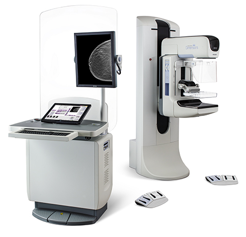
In 2011, Hologic was the first manufacturer to receive US FDA clearance for a tomosynthesis breast imaging system. From a clinical research standpoint, that also means “the 100 clinical studies that exist to date highlighting the benefits of tomosynthesis were all conducted on our system,” says Pete Valenti, Hologic’s Division President, Breast and Skeletal Health Solutions.
“We hope the TMIST trial offers similar results to the other clinical studies that have been conducted to date highlighting the benefits of tomosynthesis,” he adds.
In particular, Valenti points out the research letter published in the April 26, 2016, issue of JAMA by Dr. Elizabeth Rafferty, which concluded that Hologic’s Genius™ 3D MAMMOGRAPHY™ exams reduce recall rates and increase invasive cancer detection in women with both dense and non-dense breasts—although he finds the improvements in women with heterogeneously dense breasts to be most important.
“According to Dr. Rafferty’s research letter, improvements in both recall rate reduction and invasive cancer detection using tomosynthesis were greatest for women with heterogeneously dense breasts, with a 50% increase in invasive cancer detection and a simultaneous 14% reduction in recall rates,” he adds.
Since many women are paying for tomosynthesis exams out-of-pocket due to limited third-party reimbursement, Valenti believes that spreading the word of promising results like Dr. Rafferty’s is a critical task for Hologic.
“In fact, we’re pleased that in early August, the National Comprehensive Cancer Network included tomosynthesis for the first time in its recommended breast cancer screening guidelines,” Valenti says.
Valenti also says that recently published research literature supports comparable benefits of Hologic’s Low Dose 3D MAMMOGRAPHY exam with C-View software at a dose below the established guidelines and the company’s standard Genius 3D MAMMOGRAPHY exam. He also points out the company’s 2016 introduction of Affirm prone breast biopsy system, reportedly the world’s first and only 2D/3D dedicated prone breast biopsy system.
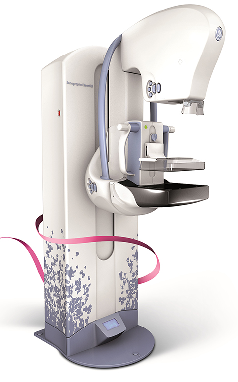
GE Healthcare, meanwhile, is offering its installed base customers the latest in 3D mammography through an upgrade to SenoClaire. According to Colleen Healy, Breast Imaging Marketing Manager, the company is also working to meet clinical needs through software features that provide enhanced reading and workflow capabilities, as well as limiting the dose delivered to patients.
“With no increase in radiation dose compared to a standard 2D mammogram, SenoClaire uses a ‘step-and-shoot’ method that reduces blur and increases image sharpness, and ASiR iterative reconstruction technology for improved image quality,” Healy explains.
Healy also expects TMIST to demonstrate that tomosynthesis should replace digital mammography as the preferred screening method. Yet, she too knows that reimbursement is a hurdle.
“Minimizing economic barriers to accessible medical services tends to increase demand, she says. “One key provision of the 2010 Patient Protection and Affordable Care Act (PPACA) was to eliminate out-of-pocket costs for USPSTF- approved preventive services, such as screening mammography.”
While CMS announced widespread coverage of digital breast tomosynthesis, effective January 2015, Healy says it is currently estimated that only 35 to 40% of women eligible for tomosynthesis screening have some form of insurance coverage for the exam.
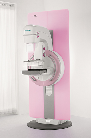
Siemens Healthineers recently received FDA clearance for stand-alone, 3D digital breast tomosynthesis screening on the Mammomat Inspiration with Tomosynthesis Option, making it the first such system with this distinction according to Jennifer Okken, Director or Mammography Products at Siemens.
According to Okken, there are two key technology differentiators of the Mammomat Inspiration with Tomosynthesis Option. “We have a wide angulation of 50 degrees, doubling the amount of data our users can acquire with one sweep. The second is our dose reduction; with the addition of our PRIME technology, our customers can reduce the patient dose by up to 30%.”
While Okken acknowledges this technology is a big step forward, there remains a need to demonstrate the clinical efficacy of the technology.
“We’re seeing an increase in the detection of cancers with tomosynthesis, and we’re hoping TMIST will demonstrate that it is superior to 2D mammography and can replace it as the standard of care,” Okken says.
While the lack of widespread reimbursement for tomosynthesis screening exams is a hurdle for wider adoption of the technology, Okken is encouraged by recent laws in Pennsylvania and Illinois that mandate coverage. Siemens, she says, is committed to educating providers and users on tomosynthesis so they can further inform third-party payers in their regions about the clinical—and potential economic—benefits of tomosynthesis.
Okken also says that for patients classified as BI-RADS 4 and 5, it is likely they will undergo both tomosynthesis and ultrasound breast screening exams. While this is not yet clearly determined, it is yet another area where innovators and manufacturers can collaborate with clinicians to define how these technologies can and should be used.
Reference
- Shapiro S, Venet W, Strax P, Venet L, Roeser R. Ten- to fourteen-year effect of screening on breast cancer mortality. J Natl Cancer Inst. 1982 Aug;69(2):349-355.
Citation
Massat M. Updates in breast imaging: TMIST, guidelines and tomosynthesis technology. Appl Radiol. 2016; (9):33-36.
September 5, 2016