White light scanner - Alternative to CT for assessment of pediatric chest deformities?
A handheld white light scanner shows potential to replace the use of radiographs and CT scans to measure and monitor the degree and severity of chest deformities in growing children. Physicians at Ann and Robert H. Lurie Children’s Hospital of Chicago who have been evaluating accuracy of the technology reported preliminary findings of their pilot study at the annual meeting of the American Pediatric Surgical Association (APSA) held in San Diego in May.
Principal investigator Marletta Reynolds, MD, surgeon-in-chief at the hospital and professor of surgery at Northwestern University Feinberg School of Medicine, reported that the radiation-free scanner has accurately and reliably captured abnormal chest shape in detailed 3D format of 53 patients with sunken chests. Pectus excavatum is the most common of upper body defects, occurring in one of 300 to 400 children.
 Pediatric patients with pectus excavatum generally have regular evaluations to monitor how their chest deformity changes in time as they grow or following surgery. Surgical correction of chest malformations is generally postponed until adolescence. However, regular monitoring is essential for all children with the condition because even mild chest defects can cause neck and back pain. Such pain can reduce a child’s cardiovascular endurance and exercise capacity. Monitoring was originally done through visual evaluation and measurement by a physician, but was replaced by X-ray exams and subsequently, by CT scans. CT imaging provides excellent visualization, but at the cost of repeated exposure to high radiation dose.
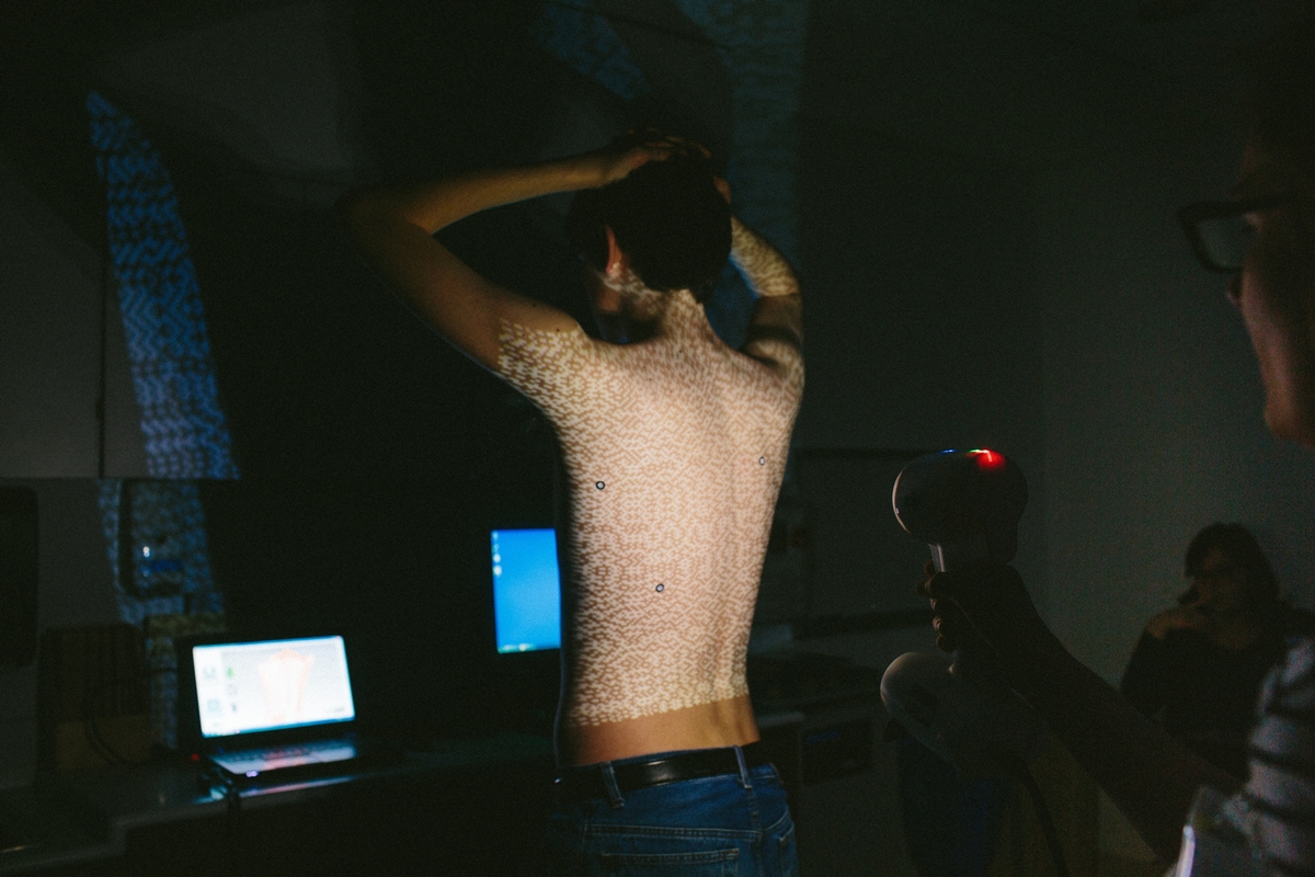
Dr. Reynolds and colleagues launched a prospective observational study in 2015. The objective of the study has been to assess the precision and accuracy of the Rodin M4D White Light Scanner in preoperative evaluation of patients with pectus excavatum. Measurements of the scanner are being compared with the current standard of care at Lurie Children’s Hospital, and when appropriate, a CT scan derived Haller index for assessing the progression of the deformity in these patients.
White-light scanning has been used in eye imaging for many years, but the researchers believe that their study is the first time that the technology has been adapted for visualizing chest malformations. They hope that the final results of this study can influence how a child with pectus excavatum is followed over time without the need for x-ray exposure and in assessing post operative correction over time.
In addition to minimizing or eliminating radiation exposure to vulnerable children, white light scanning offers the advantage of being a much less costly technology to use (purchase price is approximately $35,000) and is compact and lightweight, at less than two pounds. Its price and mobility make the researcher team hopeful that the scanner can be used in countries or regions where advanced imaging is not available or is not affordable. “Children everywhere deserve to have the same access to safe and reliable imaging technology, and we hope that our approach will provide a much-needed cheapk, quick and safe diagnostic and monitoring tool for kids in economically disadvantaged areas, “ said Fred Hebal, MD, the first author on the study and researcher in the Division of Pediatric Surgery.