Tomosynthesis provides a definitive picture for improved breast cancer diagnosis
Images


Case Summary
A 35-year-old female presented with a new lump at the left nipple, noted for about one week. She had no other palpable masses. The patient’s paternal aunt and paternal great aunt had breast cancer. A 2-dimensional + 3-dimensional (breast tomosynthesis) exam was performed on a Hologic Selenia Dimensions system with nipple markers in place and a triangular marker over the palpable lump.
Diagnosis
T1c, N1, M0 stage 2 left breast cancer with 2 separate primaries:
- 1.3-cm infiltrating ductal carcinoma left UOQ, grade 3, estrogen and progesterone receptors > 90% positive. HER-2/neu negative. One out of 7 lymph nodes positive at 1.2 mm (micromets).
- The second tumor 7 cm from the palpable retrorareolar tumor: infiltrating ductal carcinoma 1.3 cm, grade 1 ER 50% and PR > 90%. HER-2/neu negative.
Findings
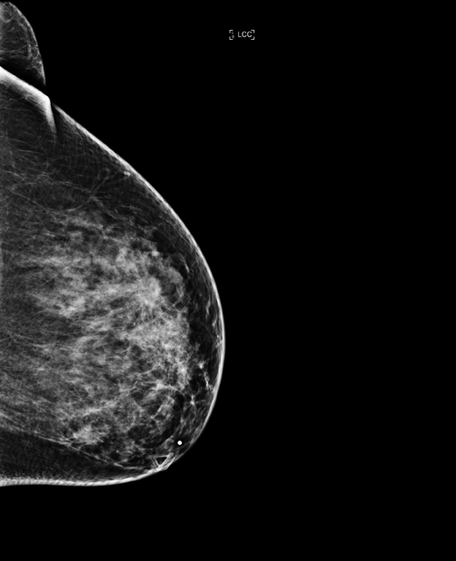
The patient’s 2-dimensional mammogram reveals dense breasts with multiple areas of increased density and mild distortions throughout both breasts, with no clear correlates (Figure 1). The 2-dimensional mammogram showed a triangle marking a palpable retroareolar mass. The retroareolar mass was not well seen with the 2-dimensional mammogram.
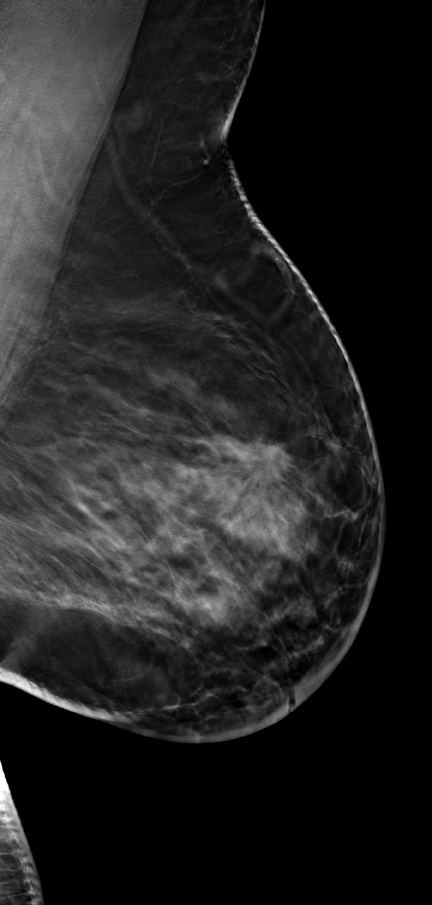
The 3-dimensional breast tomosynthesis images as seen in Figure 2 clearly showed a spiculated mass in the left upper outer quadrant (UOQ): CC slice 36, mediolateral oblique (MLO) slice 40, measuring about 1.6 cm.
Corresponding to the palpable area at the nipple, ultrasound revealed a 1.1-cm thickening. Ultrasound also showed a 1.7-cm hypoechoic mass at 2 o’clock left breast, with irregular margins and slightly increased vascularity.
Discussion
Breast tomosynthesis is a new method for breast cancer screening and diagnosis. Unlike prior-generation mammography systems, which generate 2-dimensional images, breast tomosynthesis produces 3-dimensional images that are intended to reveal the inner architecture of the breast, free from the distortion typically caused by tissue shadowing or density.
In clinical studies Hologic submitted to the FDA, radiologists reading 2-dimensional + 3-dimensional mammography (breast tomosynthesis) compared to 2-dimensional mammography alone demonstrated superior clinical performance in specificity, the confidence to rule out breast cancer without recalling the patient for further study, and also demonstrated improved sensitivity, the proportion of mammograms which include breast cancers that were correctly diagnosed. Based on pooled multi-reader receiver operating characteristic (ROC) analysis, Hologic’s clinical trials reviewed by the FDA showed the potential to increase cancer detection by up to 16% and the potential to reduce recall rate by up to 40% when using combo-mode (2-dimensional + 3-dimensional breast tomosynthesis) compared with 2-dimensional mammography alone.1
In the setting of dense breasts with many potential areas of concern on the 2-dimensional mammogram, the breast tomosynthesis study was the definitive tool in this patient’s diagnosis. The sonography finding at the area of palpable concern was mild, and in the absence of the strong tomosynthesis result, it is possible that the palpable nipple lesion could have been “downplayed” and she might not have received the immediate care she needed (biopsies of both lesions were done the following day).
The 3-dimensional (breast tomosynthesis) images obtained at the same time as the 2-dimensional images and under the same compression clearly demonstrated a mass with spiculated margins allowing very accurate localization in the UOQ and size determination. Based on the breast tomosynthesis information, we were able to very confidently predict that this would be a malignancy. Additionally, the 3-dimensional (breast tomosynthesis) imaging easily proved that other similar areas of asymmetry in the patient’s dense busy breast were due to superimposed tissues rather than additional masses, particularly medially on the left CC view.
The patient had a bilateral mastectomy. The right breast was negative for malignancy. The patient is undergoing chemotherapy at this time.
Conclusion
Three-dimensional breast tomosynthesis clearly allows better visualization of tissues in dense breasts and accurate localization for ultrasound and biopsy studies or for localization for additional mammographic imaging. (For example, knowing that calcifications are located medially so as not to waste time imaging the lateral aspect of the breast). Breast tomosynthesis also allowed us to better see or identify internal calcifications within masses.
In this case and other tomosynthesis cases, we have been able to definitively identify additional unsuspected lesions when cancers are multifocal or multicentric, which could help decrease the need for second-look ultrasound and biopsy after MRI.
- The Hologic Selenia Dimensions clinical studies presented to the FDA as part of Hologic’s PMA (P080003) submission that compared Hologic’s Selenia Dimensions 2-dimensional + 3-dimensional tomosynthesis to Hologic’s 2D FFDM.
Related Articles
Citation
. Tomosynthesis provides a definitive picture for improved breast cancer diagnosis. Appl Radiol.
October 21, 2011